
11299964001
Roche
5-Bromo-2′-deoxy-uridine Labeling and Detection Kit II
sufficient for ≤100 tests, storage temp.:-20°C
Sinonimo/i:
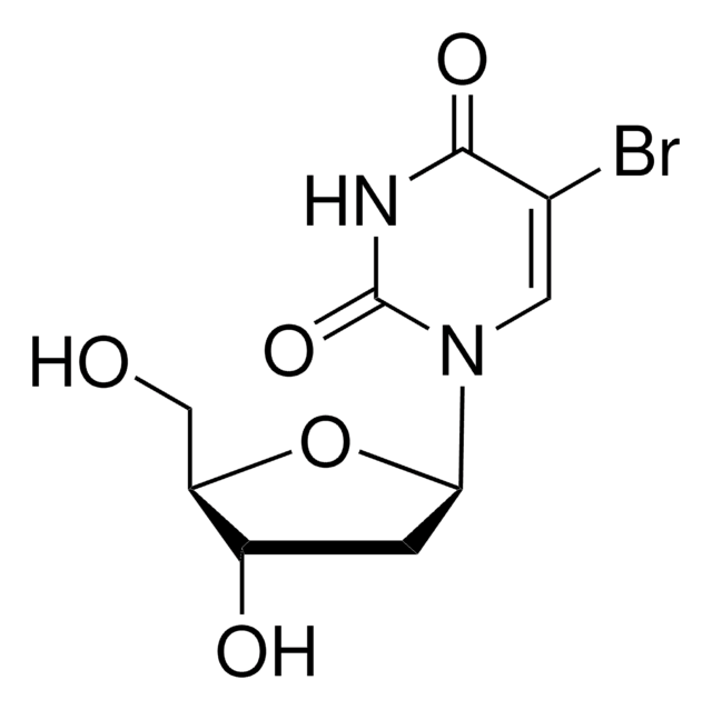
5-BrdU, 5-Bromo-2-deoxyuridine
Autenticatiper visualizzare i prezzi riservati alla tua organizzazione & contrattuali
About This Item
Codice UNSPSC:
41116133
Prodotti consigliati


impiego
sufficient for ≤100 tests
Produttore/marchio commerciale
Roche
Temperatura di conservazione
−20°C
Descrizione generale
Cell proliferation may be studied by monitoring the incorporation of a radioisotope, [ 3H]-thymidine, into cellular DNA, followed by autoradiography.
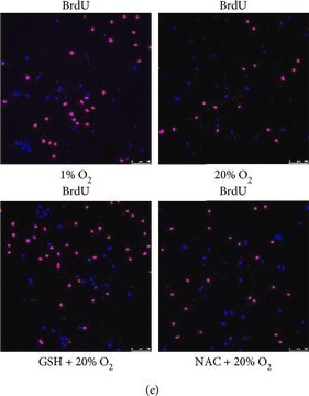
Alternatively, 5-bromo-2′-deoxy-uridine (BrdU) may be used instead of thymidine. Cells that have incorporated BrdU into DNA are easily detected using a monoclonal antibody against BrdU and an enzyme- or fluorochrome-conjugated second antibody.
Alternatively, 5-bromo-2′-deoxy-uridine (BrdU) may be used instead of thymidine. Cells that have incorporated BrdU into DNA are easily detected using a monoclonal antibody against BrdU and an enzyme- or fluorochrome-conjugated second antibody.
Immunohistocytochemical assay for the detection of 5-bromo-2′-deoxy-uridine (BrdU) incorporated into cellular DNA.
Specificità
Anti-BrdU monoclonal antibody specifically binds to 5-bromo-2′-deoxy-uridine, and shows cross-reactivity with 5-iodo-2′-deoxy-uridine (10%). Anti-BrdU shows no cross-reactivity with 5-fluoro-2′-deoxy-uridine or any endogenous cellular component, such as thymidine or uridine.
Applicazioni
5-Bromo-2′-deoxy-uridine Labeling and Detection Kit II has been used in:
- labeling of tooth roots for histology
- immunostaining of mice frontal sections
- immunofluorescence imaging of hepatocellular carcinoma sections
The kit is used for the detection of BrdU incorporated into cellular DNA by immunohistocytochemistry.
Caratteristiche e vantaggi
- Safe: No radioisotopes are used.
- Easy to perform: Follows a standard immunohistochemistry protocol.
- Sensitive: Denaturation of DNA with nucleases allows for highly sensitive detection of BrdU.
- Flexible: Allows double-labeling protocols.
Confezionamento
1 kit containing 7 components.
Principio
Samples prelabeled with BrdU are fixed with ethanol, then incubated with a monoclonal antibody to BrdU, which contains an optimized mixture of nucleases. These nucleases generate single-stranded DNA fragments that allow binding of the antibody to BrdU. Next, an alkaline phosphatase (AP)-labeled antibody to mouse immunoglobulin is added, then bound to the anti-BrdU antibody. The sample is then incubated with the AP substrate and NBT/ BCIP, which is metabolized to form a colored reaction product. The sample is evaluated using a phase-contrast microscope.
Nota sulla preparazione
Working concentration: Working concentration of the labeling reagent corresponds to the WC of the In Situ Cell Proliferation Kits.
Sample material:
Cell culture: adherent cells, suspension cells, organ or explant cultures. Frozen or paraffin-embedded tissue sections (after in vivo labeling).
Sample material:
Cell culture: adherent cells, suspension cells, organ or explant cultures. Frozen or paraffin-embedded tissue sections (after in vivo labeling).
Altre note
For life science research only. Not for use in diagnostic procedures.
Solo come componenti del kit
N° Catalogo
Descrizione
- BrdU Labeling Reagent 1,000x concentrated
- Washing Buffer concentrate 10x concentrated
- Incubation Buffer
- Anti-BrdU antibody, contains nucleases for DNA denaturation
- Anti-mouse Ig-alkaline Phosphatase antibody
- NBT
- BCIP


Avvertenze
Danger
Indicazioni di pericolo
Consigli di prudenza
Classi di pericolo
Acute Tox. 4 - Acute Tox. 4 Inhalation - Eye Irrit. 2 - Flam. Liq. 3 Dermal - Muta. 1B - Repr. 1B - Skin Sens. 1
Codice della classe di stoccaggio
3 - Flammable liquids
Classe di pericolosità dell'acqua (WGK)
WGK 2
Punto d’infiammabilità (°F)
136.4 °F
Punto d’infiammabilità (°C)
58 °C
Scegli una delle versioni più recenti:
Possiedi già questo prodotto?
I documenti relativi ai prodotti acquistati recentemente sono disponibili nell’Archivio dei documenti.
I clienti hanno visto anche

A Lecointe et al.
Cytotechnology, 65(5), 705-724 (2013-06-13)
Cell cultures from reef-building scleractinian corals are being developed to study the response of these ecologically important organisms to environmental stress and diseases. Despite the importance of cell division to support propagation, cell proliferation in polyps and in vitro is
Pten regulates neural crest proliferation and differentiation during mouse craniofacial development
Yang T, et al.
Developmental Dynamics, 247(2), 304-314 (2018)
CXCL14 and MCP1 are potent trophic factors associated with cell migration and angiogenesis leading to higher regenerative potential of dental pulp side population cells
Hayashi Y, et al.
Stem Cell Research & Therapy, 6(1), 111-111 (2015)
L J Backman et al.
Scandinavian journal of medicine & science in sports, 23(6), 687-696 (2012-02-02)
The histopathology of tendons with painful tendinopathy is often tendinosis, a fibrosis-like condition of unclear pathogenesis characterized by tissue changes including hypercellularity. The primary tendon cells (tenocytes) have been shown to express adrenoreceptors (mainly alpha-2A) as well as markers of
Hitoshi Ikeda et al.
Journal of lipid research, 50(3), 556-564 (2008-10-29)
Sphingosine 1-phosphate (S1P), a bioactive lipid mediator, stimulates proliferation and contractility in hepatic stellate cells, the principal matrix-producing cells in the liver, and inhibits proliferation via S1P receptor 2 (S1P(2)) in hepatocytes in rats in vitro. A potential role of
Articoli
Cell based assays for cell proliferation (BrdU, MTT, WST1), cell viability and cytotoxicity experiments for applications in cancer, neuroscience and stem cell research.
Il team dei nostri ricercatori vanta grande esperienza in tutte le aree della ricerca quali Life Science, scienza dei materiali, sintesi chimica, cromatografia, discipline analitiche, ecc..
Contatta l'Assistenza Tecnica.