Passa al contenuto
Merck
Tutte le immagini(1)

Documenti fondamentali

MABT1502

Sigma-Aldrich

Anti-Integrin beta-1 Antibody, clone 102DF5

clone 102DF5, from mouse

Sinonimo/i:

Fibronectin receptor subunit beta, Glycoprotein IIa, GPIIA, VLA-4 subunit beta, CD29

Autenticatiper visualizzare i prezzi riservati alla tua organizzazione & contrattuali


About This Item

Codice UNSPSC:
12352203
eCl@ss:
32160702
NACRES:
NA.41

Origine biologica

mouse

Forma dell’anticorpo

purified antibody

Tipo di anticorpo

primary antibodies

Clone

102DF5, monoclonal

Reattività contro le specie

human

Confezionamento

antibody small pack of 25 μg

tecniche

ELISA: suitable
flow cytometry: suitable
immunofluorescence: suitable
immunohistochemistry: suitable (paraffin)
immunoprecipitation (IP): suitable
western blot: suitable

Isotipo

IgG1κ

N° accesso NCBI

N° accesso UniProt

modifica post-traduzionali bersaglio

unmodified

Informazioni sul gene

human ... ITGB1(3688)

Descrizione generale

Integrin beta-1 (UniProt: P05556; also known as Fibronectin receptor subunit beta, Glycoprotein IIa, GPIIA, VLA-4 subunit beta, CD29) is encoded by the ITGB1 (also known as FNRB, MDF2, MSK12) gene (Gene ID: 3688) in human. Integrins are heterodimeric integral membrane proteins composed of an alpha subunit and a beta subunit that function in cell surface adhesion and signaling. Integrin beta-1 is a single-pass type I membrane protein that can associate with one of the multiple alpha subunits to serve as a receptor for fibronectin and other extracellular matrix proteins. Integrin beta-1 integrin recognizes the sequence R-G-D in a wide array of ligands. Five isoforms of integrin beta-1 have been reported that are produced by alternative splicing. Integrin beta-1 is synthesized with a signal peptide (aa 1-20) that is subsequently cleaved off. The mature form has an extracellular domain (aa 21-728), a helical domain (aa 729-751) and a cytoplasmic region (aa 752-798). Isoform 1 of integrin beta-1 is widely expressed and other isoforms are generally co-expressed with a more restricted distribution. Isoform 2 is expressed in skin, liver, skeletal muscle, cardiac muscle, placenta, umbilical vein endothelial cells, neuroblastoma cells, lymphoma cells, hepatoma cells and astrocytoma cells. Isoform 3 and isoform 4 are expressed in muscle, kidney, liver, placenta, cervical epithelium, umbilical vein endothelial cells, fibroblast cells, embryonal kidney cells, platelets and several blood cell lines. Isoform 4 is selectively expressed in peripheral T-cells and isoform 5 is expressed specifically in striated muscle (skeletal and cardiac muscle). Isoform 2 is reported to interfere with isoform 1 resulting in a dominant negative effect on cell adhesion and migration (in vitro).

Specificità

Clone 102DF5 specifically detects integrin beta-1 subunit in human cells.

Immunogeno

Tissue extract from human myometrium.

Applicazioni

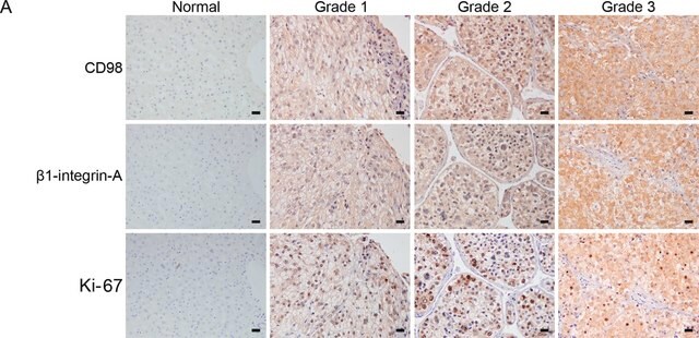
Anti-Integrin beta-1, clone 102DF5, Cat. No. MABT1502, is a mouse monoclonal antibody that detects Integrin beta-1 and has been tested for use in ELISA, Flow Cytometry, Immunofluorescence, Immunohistochemistry (Paraffin), Immunoprecipitation, and Western Blotting.
Flow Cytometry Analysis: A representative lot detected Integrin beta-1 in Flow Cytometry applications (Belkin, A.M., et. al. (1997). J Cell Biol. 139(6):1583-95).

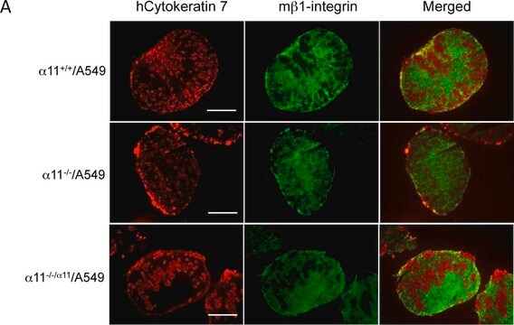
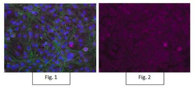
Immunofluorescence Analysis: A representative lot detected Integrin beta-1 in Immunofluorescence applications (Cartier-Michaud, A., et. al. (2012). PLoS One. 7(2):e32204; Balzac, F., et. al. (1993). J Cell Biol. 121(1):171-8; Lin, Y.N., et. al. (2015). Oncotarget. 6(21):18577-89).

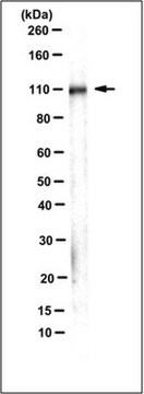
Western Blotting Analysis: A representative lot detected Integrin beta-1 in Western Blotting applications (Balzac, F., et. al. (1993). J Cell Biol. 121(1):171-8).

ELISA Analysis: A representative lot detected Integrin beta-1 in ELISA applications (Jovanovic, M., et. al. (2010). Act Histochem. 112(1):34-41).

Immunoprecipitation Analysis: A representative lot immunoprecipitated Integrin beta-1 in Immunoprecipitation applications (Balzac, F., et. al. (1993). J Cell Biol. 121(1):171-8).
Research Category
Cell Structure

Qualità

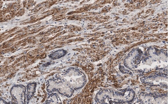
Evaluated by Immunohistochemistry (Paraffin) in human uterus and human kidney tissue sections.

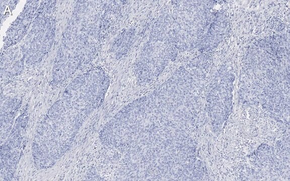
Immunohistochemistry (Paraffin) Analysis: A 1:250 dilution of this antibody detected Integrin beta-1 in human uterus and human kidney tissue sections.

Descrizione del bersaglio

~150 kDa observed; 88.42 kDa calculated. Uncharacterized bands may be observed in some lysate(s).

Stato fisico

Format: Purified
Protein G purified
Purified mouse monoclonal antibody IgG1 in buffer containing 0.1 M Tris-Glycine (pH 7.4), 150 mM NaCl with 0.05% sodium azide.

Stoccaggio e stabilità

Stable for 1 year at 2-8°C from date of receipt.

Altre note

Concentration: Please refer to lot specific datasheet.

Esclusione di responsabilità

Unless otherwise stated in our catalog or other company documentation accompanying the product(s), our products are intended for research use only and are not to be used for any other purpose, which includes but is not limited to, unauthorized commercial uses, in vitro diagnostic uses, ex vivo or in vivo therapeutic uses or any type of consumption or application to humans or animals.

Non trovi il prodotto giusto?  

Prova il nostro Motore di ricerca dei prodotti.

Codice della classe di stoccaggio

12 - Non Combustible Liquids

Classe di pericolosità dell'acqua (WGK)

WGK 1


Certificati d'analisi (COA)

Cerca il Certificati d'analisi (COA) digitando il numero di lotto/batch corrispondente. I numeri di lotto o di batch sono stampati sull'etichetta dei prodotti dopo la parola ‘Lotto’ o ‘Batch’.

Possiedi già questo prodotto?

I documenti relativi ai prodotti acquistati recentemente sono disponibili nell’Archivio dei documenti.

Visita l’Archivio dei documenti

Il team dei nostri ricercatori vanta grande esperienza in tutte le aree della ricerca quali Life Science, scienza dei materiali, sintesi chimica, cromatografia, discipline analitiche, ecc..

Contatta l'Assistenza Tecnica.