MABC97
Anti-S1PR4/EDG6 Antibody, clone 16B9.1
clone 16B9.1, from mouse
别名:
Sphingosine 1-phosphate receptor 4, S1P receptor 4, S1P4, Endothelial differentiation G-protein coupled receptor 6, Sphingosine 1-phosphate receptor Edg-6, S1P receptor Edg-6
登录查看公司和协议定价
所有图片(2)
About This Item
分類程式碼代碼:
12352203
eCl@ss:
32160702
NACRES:
NA.41
推荐产品
生物源
mouse
品質等級
抗體表格
purified immunoglobulin
抗體產品種類
primary antibodies
無性繁殖
16B9.1, monoclonal
物種活性
rat, human
技術
immunohistochemistry: suitable
western blot: suitable
同型
IgMκ
NCBI登錄號
UniProt登錄號
運輸包裝
wet ice
目標翻譯後修改
unmodified
基因資訊
human ... S1PR4(8698)
一般說明
Sphingosine 1-phosphate receptor 4 (S1PR4), otherwise known as EDG6, is present, in varying expression levels, in the adult lymph node, bone marrow, and thymus, as well as fetal liver, lung, and thymus. It is activated by the lysosphingolipid sphingosine 1-phosphate, and is coupled to Gi and G12/13 G proteins and via their various effector molecules, SIP4 regulates cell proliferation, differentiation, motility, and migration.
免疫原
GST-tagged recombinant protein corresponding to human S1PR4/EDG6.
應用
Detect S1PR4/EDG6 using this Anti-S1PR4/EDG6 Antibody, clone 16B9.1 validated for use in Western Blotting & IHC.
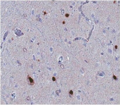
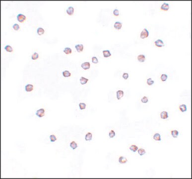
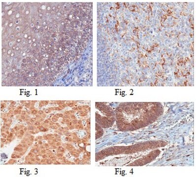
Immunohistochemistry Analysis: A 1:300 dilution from a representative lot detected S1PR4/EDG6 in human red bone marrow and human tonsil tissues.
品質
Evaluated by Western Blot Analysis in rat spleen tissue lysate.
Western Blot Analysis: 1 µg/mL of this antibody detected S1PR4/EDG6 in 10 µg of rat spleen tissue lysate.
Western Blot Analysis: 1 µg/mL of this antibody detected S1PR4/EDG6 in 10 µg of rat spleen tissue lysate.
標靶描述
~42 kDa observed
外觀
Format: Purified
其他說明
Concentration: Please refer to the Certificate of Analysis for the lot-specific concentration.
未找到合适的产品?
试试我们的产品选型工具.
儲存類別代碼
10 - Combustible liquids
水污染物質分類(WGK)
WGK 2
閃點(°F)
Not applicable
閃點(°C)
Not applicable
Qun Zhu et al.
The FEBS journal, 281(12), 2861-2870 (2014-05-07)
It has been reported that the effect of inflammatory cytokines on β-cell destruction in type 1 diabetes is concentration-dependent. However, the underlying mechanisms remain unclear. In the present study, we found that a high concentration of cytokines promoted apoptosis in
我们的科学家团队拥有各种研究领域经验,包括生命科学、材料科学、化学合成、色谱、分析及许多其他领域.
联系技术服务部门