推荐产品
生物源
rabbit
品質等級
抗體表格
purified immunoglobulin
抗體產品種類
primary antibodies
無性繁殖
polyclonal
物種活性
monkey, human, mouse
製造商/商標名
Chemicon®
技術
immunohistochemistry: suitable (paraffin)
NCBI登錄號
UniProt登錄號
運輸包裝
wet ice
目標翻譯後修改
unmodified
基因資訊
human ... OPN1LW(5956)
一般說明
人类的全部颜色辨别是基于三种视锥细胞感光机制的存在和功能。 每种视锥类型都具有由11-顺式视黄醛和独特的视蛋白组成的光敏色素-蛋白复合物,其在光谱的短波长(S视锥,峰值灵敏度约为420nm),中波长(M视锥,峰值灵敏度约为530nm,具有多态性;Winderckx et al., 1993; Neitz & Neitz, 1998)和长波长(L视锥,峰值灵敏度约为560nm,具有多态性;Neitz & Jacobs, 1990)下都具有灵敏度。 全部三种视蛋白是具有七个跨膜区域的跨膜蛋白。 三种类型的视锥蛋白和视杆感光视紫红质基因的基因似乎是同源的,具有不同的保守程度。 最强的保守性在X染色体上的中波长(绿色)和长(红色)波长敏感色素之间,表明相对较新的复制/发散事件(Nathans, 1989; Nathans et al., 1992)。 S视锥(蓝色)视蛋白位于7号染色体上,似乎与视紫红质具有更强的保守性。 人的视锥细胞光受体分布主要由M和L视锥色素控制。
特異性
可识别视蛋白,蓝色。
免疫原
表位:蓝色
重组的人蓝色视蛋白。
應用
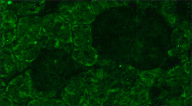
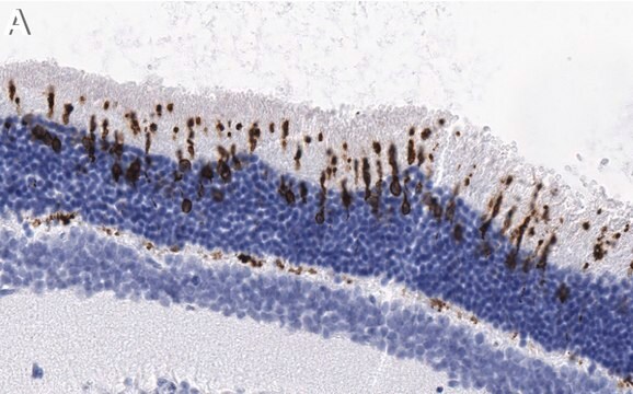
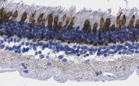
免疫组化:在福尔马林固定,石蜡包埋的小鼠视网膜组织上1:200-1:300。抗原修复方法推荐是采用蒸汽加热的HIER;其他固定和回收方法未经测试。
最佳工作稀释度必须由最终用户进行确定。
最佳工作稀释度必须由最终用户进行确定。
研究子类别
感官 & PNS
感官 & PNS
研究类别
神经科学
神经科学
该蓝色抗视蛋白抗体经过验证可用于IH(P)中检测视蛋白。
外觀
形式:纯化
纯化免疫球蛋白PBS{0.02 M磷酸盐,0.25 M NaCl,pH值7.6},含0.1%叠氮化钠作为防腐剂
纯化的蛋白 A
儲存和穩定性
自发运之日起,在 2–8°C 条件下可稳定保存 1 年
分析報告
对照
视网膜
视网膜
法律資訊
CHEMICON is a registered trademark of Merck KGaA, Darmstadt, Germany
免責聲明
除非我们的产品目录或产品附带的其他公司文档另有说明,否则我们的产品仅供研究使用,不得用于任何其他目的,包括但不限于未经授权的商业用途、体外诊断用途、离体或体内治疗用途或任何类型的消费或应用于人类或动物。
未找到合适的产品?
试试我们的产品选型工具.
儲存類別代碼
12 - Non Combustible Liquids
水污染物質分類(WGK)
WGK 1
閃點(°F)
Not applicable
閃點(°C)
Not applicable
Separate blue and green cone networks in the mammalian retina.
Li, Wei and DeVries, Steven H
Nature Neuroscience, 7, 751-756 (2004)
NeuroD1 regulates expression of thyroid hormone receptor 2 and cone opsins in the developing mouse retina.
Liu, H; Etter, P; Hayes, S; Jones, I; Nelson, B; Hartman, B; Forrest, D; Reh, TA
The Journal of Neuroscience null
Transcriptional profile analysis of RPGRORF15 frameshift mutation identifies novel genes associated with retinal degeneration.
Genini, S; Zangerl, B; Slavik, J; Acland, GM; Beltran, WA; Aguirre, GD
Investigative Ophthalmology & Visual Science null
The electroretinogram (ERG) of a diurnal cone-rich laboratory rodent, the Nile grass rat (Arvicanthis niloticus).
Gilmour, Gregory S, et al.
Vision Research, 48, 2723-2731 (2008)
L P Morin et al.
Neuroscience, 199, 213-224 (2011-10-12)
Four studies were performed to further clarify the contribution of rod/cone and intrinsically photoreceptive retinal ganglion cells to measures of entrainment, dark preference, light-induced locomotor suppression and photosomnolence. Wild type (WT), retinally degenerate (rd/rd), and melanopsin-less (OPN4⁻/⁻) mouse strains were
我们的科学家团队拥有各种研究领域经验,包括生命科学、材料科学、化学合成、色谱、分析及许多其他领域.
联系技术服务部门


