MAB4234-C
Anti-Wilms′ Tumor Antibody, NT clone 6F-H2, Ascites Free
clone 6F-H2, from mouse
Sinónimos:
Wilms tumor protein, WT33
About This Item
Productos recomendados
origen biológico
mouse
Nivel de calidad
forma del anticuerpo
purified immunoglobulin
tipo de anticuerpo
primary antibodies
clon
6F-H2, monoclonal
reactividad de especies
mouse, human
técnicas
immunocytochemistry: suitable
immunofluorescence: suitable
immunohistochemistry: suitable (paraffin)
immunoprecipitation (IP): suitable
western blot: suitable
isotipo
IgG1κ
Nº de acceso NCBI
Nº de acceso UniProt
Condiciones de envío
wet ice
modificación del objetivo postraduccional
unmodified
Información sobre el gen
human ... WT1(7490)
Descripción general
Aplicación
Immunoprecipitation Analysis: A representative lot co-immunoprecipitated CRE-binding protein/CBP together with Wilms tumor protein WT1 from the lysate of a T-SV40 immortalized human glomerular epithelial cell (HGEC) line (Drossopoulou, G.I., et al. (2009). Am. J. Physiol. Renal Physiol. 297(3):F594-F603).
Western Blotting Analysis: A representative lot detected Wilms tumor protein WT1 in the CRE-binding protein/CBP immunoprecipitate obtained from the lysate of a T-SV40 immortalized human glomerular epithelial cell (HGEC) line (Drossopoulou, G.I., et al. (2009). Am. J. Physiol. Renal Physiol. 297(3):F594-F603).
Western Blotting Analysis: A representative lot detected Wilms tumor protein WT1 in lysates from mouse E15.5 embryonic kidney and human melanoma cell lines A375, SK-MEL-28, and WM-266-4 (Wagner, N., et al. (2008). Pflugers Arch. Eur. J. Physiol. 455(5):839-847).
Immunocytochemistry Analysis: A representative lot immunostained the nucleus of methanol-fixed human melanoma A375 cells by fluorescent immunocytochemistry (Wagner, N., et al. (2008). Pflugers Arch. Eur. J. Physiol. 455(5):839-847).
Immunofluorescence Analysis: A representative lot immunostained the PCNA-positive nuclei of proliferating cells in formalin-fixed, paraffin-embedded human melanoma tissue sections by fluorescent immunohistochemistry (Wagner, N., et al. (2008). Pflugers Arch. Eur. J. Physiol. 455(5):839-847).
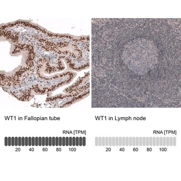
Immunohistochemistry Analysis: A representative lot immunostained glomeruli in formalin-fixed, paraffin-embedded normal human kidney and Wilms′ tumor sections (Wagner, N., et al. (2008). Pediatr. Nephrol. 23(9):1445-1453).
Immunohistochemistry Analysis: A representative lot detected vascular WT1 expression in 95% of 113 paraffin-embedded tumour tissues of various types. In most cases, nuclear WT1 staining of endothelial cells was seen (Wagner, N., et al. (2008). Oncogene. 27(26):3662-3672).
Immunohistochemistry Analysis: A representative lot immunostained the nucleus of perifollicular fibroblasts at the hair follicle in formalin-fixed, paraffin-embedded normal human skin sections. Most common melanocytic nevi do not express WT1, whereas Spitz nevi and dysplastic nevi show cytoplasmic WT1 staining. (Wagner, N., et al. (2008). Pflugers Arch. Eur. J. Physiol. 455(5):839-847).
Calidad
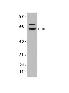
Western Blotting Analysis: 1.0 µg/mL of this antibody detected Wilms tumor protein in 10 µg of Jurkat cell lysate.
Descripción de destino
Forma física
Otras notas
¿No encuentra el producto adecuado?
Pruebe nuestro Herramienta de selección de productos.
Opcional
Código de clase de almacenamiento
12 - Non Combustible Liquids
Clase de riesgo para el agua (WGK)
WGK 1
Punto de inflamabilidad (°F)
Not applicable
Punto de inflamabilidad (°C)
Not applicable
Certificados de análisis (COA)
Busque Certificados de análisis (COA) introduciendo el número de lote del producto. Los números de lote se encuentran en la etiqueta del producto después de las palabras «Lot» o «Batch»
¿Ya tiene este producto?
Encuentre la documentación para los productos que ha comprado recientemente en la Biblioteca de documentos.
Nuestro equipo de científicos tiene experiencia en todas las áreas de investigación: Ciencias de la vida, Ciencia de los materiales, Síntesis química, Cromatografía, Analítica y muchas otras.
Póngase en contacto con el Servicio técnico
