Saltar al contenido
Merck
Todas las fotos(1)

Key Documents

05-753

Sigma-Aldrich

Anti-WT1 Antibody, clone 6F-H2

clone 6F-H2, Upstate®, from mouse

Sinónimos:

Anti-AWT1, Anti-GUD, Anti-NPHS4, Anti-WAGR, Anti-WIT-2, Anti-WT-1, Anti-WT33

Iniciar sesiónpara Ver la Fijación de precios por contrato y de la organización


About This Item

Código UNSPSC:
12352203
eCl@ss:
32160702
NACRES:
NA.41

origen biológico

mouse

Nivel de calidad

forma del anticuerpo

purified immunoglobulin

tipo de anticuerpo

primary antibodies

clon

6F-H2, monoclonal

reactividad de especies

human

fabricante / nombre comercial

Upstate®

técnicas

immunocytochemistry: suitable
immunohistochemistry: suitable
western blot: suitable

isotipo

IgG1

Nº de acceso NCBI

Nº de acceso UniProt

Condiciones de envío

wet ice

modificación del objetivo postraduccional

unmodified

Información sobre el gen

human ... WT1(7490)

Especificidad

WT1

Inmunógeno

6His-tagged fusion protein corresponding to residues 1-181 of human WT1 (Wilms tumor)

Aplicación

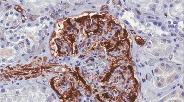
Anti-WT1 Antibody, clone 6F-H2 is a Mouse Monoclonal Antibody for detection of WT1 also known as Wilms′ Tumor & has been tested in WB, ICC & IHC.

Calidad

routinely evaluated by immunoblot on RIPA lysates from Jurkat and Raji cells

Descripción de destino

55-60kDa

Forma física

Format: Purified

Información legal

UPSTATE is a registered trademark of Merck KGaA, Darmstadt, Germany

¿No encuentra el producto adecuado?  

Pruebe nuestro Herramienta de selección de productos.

Opcional

Código de clase de almacenamiento

10 - Combustible liquids

Clase de riesgo para el agua (WGK)

WGK 1


Certificados de análisis (COA)

Busque Certificados de análisis (COA) introduciendo el número de lote del producto. Los números de lote se encuentran en la etiqueta del producto después de las palabras «Lot» o «Batch»

¿Ya tiene este producto?

Encuentre la documentación para los productos que ha comprado recientemente en la Biblioteca de documentos.

Visite la Librería de documentos

Toru Sakairi et al.
American journal of physiology. Renal physiology, 298(3), F557-F567 (2009-12-04)
Evidence suggests that loss of podocytes into urine contributes to development of glomerular diseases; shed podocytes are frequently viable and proliferate in culture conditions. To determine the phenotypic characteristics of viable urinary cells derived from human subjects, we established long-term
Zenglai Tan et al.
Scientific reports, 8(1), 16618-16618 (2018-11-11)
The kidney is a complex organ that is comprised of thousands of nephrons developing through reciprocal inductive interactions between metanephric mesenchyme (MM) and ureteric bud (UB). The MM undergoes mesenchymal to epithelial transition (MET) in response to the signaling from
Toru Sakairi et al.
American journal of physiology. Renal physiology, 299(4), F802-F809 (2010-07-30)
We transformed mouse podocytes by ectopic expression of cyclin-dependent kinase 4 (CDK4). Compared with podocytes transformed with a thermo-sensitive SV40 large T antigen mutant tsA58U19 (tsT podocytes), podocytes transformed with CDK4 (CDK4 podocytes) exhibited significantly higher expression of nephrin mRNA.
Rosalba Parenti et al.
PloS one, 9(12), e114333-e114333 (2014-12-05)
Wilms' tumor gene 1 (WT1) plays complex roles in tumorigenesis, acting as tumor suppressor gene or an oncogene depending on the cellular context. WT1 expression has been variably reported in both benign and malignant peripheral nerve sheath tumors (MPNSTs) by
Aleksandra Rak-Raszewska et al.
Developmental dynamics : an official publication of the American Association of Anatomists (2021-09-09)
Tissue organoids derived from primary cells have high potential for studying organ development and diseases in numerous organs. They recreate the morphological structure and mimic the functions of given organ while being compact in size, easy to produce, and suitable

Nuestro equipo de científicos tiene experiencia en todas las áreas de investigación: Ciencias de la vida, Ciencia de los materiales, Síntesis química, Cromatografía, Analítica y muchas otras.

Póngase en contacto con el Servicio técnico