05-665
Anti-Active-β-Catenin (Anti-ABC) Antibody, clone 8E7
clone 8E7, Upstate®, from mouse
Sinónimos:
Anti-Anti-CTNNB, Anti-Anti-EVR7, Anti-Anti-MRD19, Anti-Anti-NEDSDV, Anti-Anti-armadillo
About This Item
Productos recomendados
origen biológico
mouse
Nivel de calidad
forma del anticuerpo
purified immunoglobulin
tipo de anticuerpo
primary antibodies
clon
8E7, monoclonal
reactividad de especies
human, rat, mouse
envase
antibody small pack of 25 μg
fabricante / nombre comercial
Upstate®
técnicas
flow cytometry: suitable
immunocytochemistry: suitable
immunohistochemistry (formalin-fixed, paraffin-embedded sections): suitable
western blot: suitable
isotipo
IgG1κ
Nº de acceso NCBI
Nº de acceso UniProt
Condiciones de envío
ambient
modificación del objetivo postraduccional
unmodified
Información sobre el gen
human ... CTNNB1(1499)
mouse ... Ctnnb1(12387)
rat ... Ctnnb1(84353)
Descripción general
When β-catenin was sequenced it was found to be a member of the armadillo family of proteins. These proteins have multiple copies of the so-called armadillo repeat domain which is specialized for protein-protein binding. An increase in β-catenin production has been noted in those people who have Basal Cell Carcinoma and leads to the increase in proliferation of related tumors. When β-catenin is not associated with cadherins and α-catenin, it can interact with other proteins such as Catenin Beta Interacting Protein 1 (ICAT) and adenomatosis polyposis coli (APC).
Recent evidence suggests that β-catenin plays an important role in various aspects of liver biology including liver development (both embryonic and postnatal), liver regeneration following partial hepatectomy. HGF-induced hepatpomegaly, liver zonation, and pathogenesis of liver cancer.
Especificidad
Inmunógeno
Aplicación
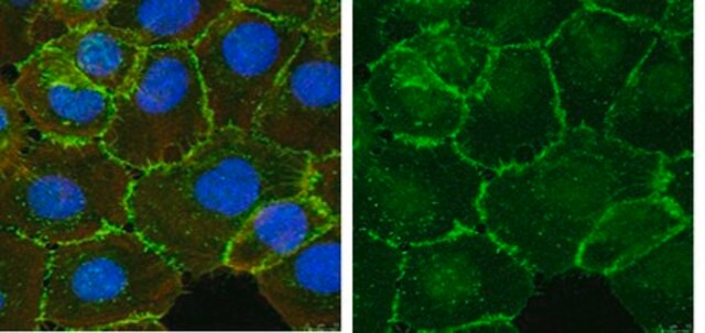
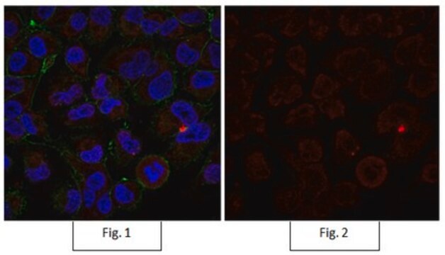
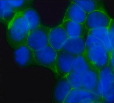
This antibody has also been reported by an independent laboratory to show positive immunostaining for beta-catenin in LiCl-treated 293T cells fixed with methanol (Staal, Frank J. T., 2002).
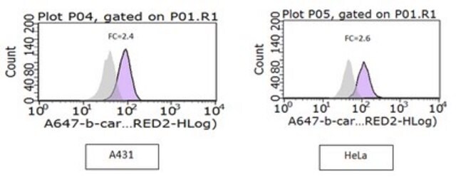
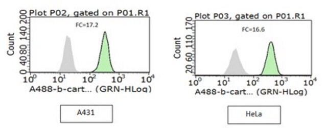
Flow Cytometry: This antibody was used in flow cytometry at an optimal 1 μg/mL concentration.
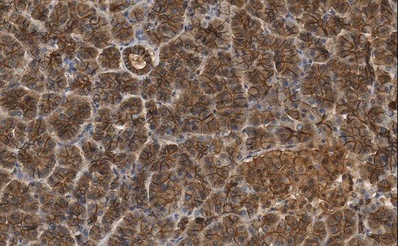
Immunohistochemistry: This antibody was used in immunohistochemistry on a colorectal carcinoma tissue array at a 1:300 dilution.
This antibody has also been reported by an independent laboratory to detect beta-catenin in mouse embryo sections (Van Noort, M., 2002).
Epigenetics & Nuclear Function
Transcription Factors
Calidad
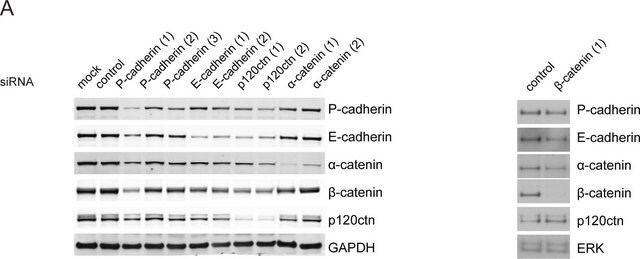
Western Blot Analysis: 0.2-2 µg/mL of this antibody detected β-catenin in RIPA lysates from A431 cells.
Descripción de destino
Forma física
Almacenamiento y estabilidad
Handling Recommendations: Upon receipt, and prior to removing the cap, centrifuge the vial and gently mix the solution.
Nota de análisis
Positive Antigen Control: Catalog #12-301, non-stimulated A431 cell lysate. Add 2.5µL of 2-mercaptoethanol/100µL of lysate and boil for 5 minutes to reduce the preparation. Load 20µg of reduced lysate per lane for minigels.
Información legal
Cláusula de descargo de responsabilidad
¿No encuentra el producto adecuado?
Pruebe nuestro Herramienta de selección de productos.
Opcional
Código de clase de almacenamiento
12 - Non Combustible Liquids
Clase de riesgo para el agua (WGK)
WGK 1
Punto de inflamabilidad (°F)
Not applicable
Punto de inflamabilidad (°C)
Not applicable
Certificados de análisis (COA)
Busque Certificados de análisis (COA) introduciendo el número de lote del producto. Los números de lote se encuentran en la etiqueta del producto después de las palabras «Lot» o «Batch»
¿Ya tiene este producto?
Encuentre la documentación para los productos que ha comprado recientemente en la Biblioteca de documentos.
Los clientes también vieron
Nuestro equipo de científicos tiene experiencia en todas las áreas de investigación: Ciencias de la vida, Ciencia de los materiales, Síntesis química, Cromatografía, Analítica y muchas otras.
Póngase en contacto con el Servicio técnico