32922
Lugol solution
according to Lugol
Sinonimo/i:
Iodine/Potassium iodide solution
About This Item
Prodotti consigliati
Forma fisica
liquid
Livello qualitativo
Qualità
according to Lugol
Densità
1.006 g/cm3 at 20 °C
applicazioni
diagnostic assay manufacturing
food and beverages
hematology
histology
Temperatura di conservazione
room temp
Stringa SMILE
[K+].[I-].II
InChI
1S/I2.HI.K/c1-2;;/h;1H;/q;;+1/p-1
XUEKKCJEQVGHQZ-UHFFFAOYSA-M
Cerchi prodotti simili? Visita Guida al confronto tra prodotti
Categorie correlate
Descrizione generale
Applicazioni
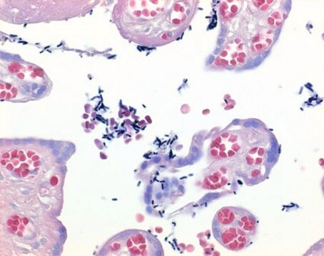
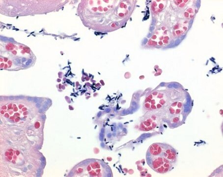
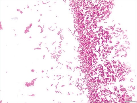
- Lugol′s solution is generally used as a mordant in Gram staining, and for fixing invertebrate blood cells and protozoa.
- It is suitable for the detection of starch in plants.
- It has been used in a study to assess narrow-band imaging without image magnification for detecting high-grade dysplasia and intramucosal esophageal squamous cell carcinoma.
- It has been used in a study to determine its benefit in aiding the detection, presence, and spread of small squamous cell carcinomas of the esophagus.
Azioni biochim/fisiol
Caratteristiche e vantaggi
- Ready-to-use solution.
Principio
Codice della classe di stoccaggio
10 - Combustible liquids
Classe di pericolosità dell'acqua (WGK)
WGK 2
Punto d’infiammabilità (°F)
Not applicable
Punto d’infiammabilità (°C)
Not applicable
Dispositivi di protezione individuale
Eyeshields, Gloves, multi-purpose combination respirator cartridge (US)
Certificati d'analisi (COA)
Cerca il Certificati d'analisi (COA) digitando il numero di lotto/batch corrispondente. I numeri di lotto o di batch sono stampati sull'etichetta dei prodotti dopo la parola ‘Lotto’ o ‘Batch’.
Possiedi già questo prodotto?
I documenti relativi ai prodotti acquistati recentemente sono disponibili nell’Archivio dei documenti.
I clienti hanno visto anche
Il team dei nostri ricercatori vanta grande esperienza in tutte le aree della ricerca quali Life Science, scienza dei materiali, sintesi chimica, cromatografia, discipline analitiche, ecc..
Contatta l'Assistenza Tecnica.


