Passa al contenuto
Merck
Tutte le immagini(3)

Documenti fondamentali

MABC28

Sigma-Aldrich

Anti-RIP3 Antibody, clone 6E6.2

clone 6E6.2, from mouse

Sinonimo/i:

Receptor-interacting serine/threonine-protein kinase 3, RIP-like protein kinase 3, Receptor-interacting protein 3, RIP-3

Autenticatiper visualizzare i prezzi riservati alla tua organizzazione & contrattuali


About This Item

Codice UNSPSC:
12352203
eCl@ss:
32160702
NACRES:
NA.41

Origine biologica

mouse

Livello qualitativo

Forma dell’anticorpo

purified immunoglobulin

Tipo di anticorpo

primary antibodies

Clone

6E6.2, monoclonal

Reattività contro le specie

human, rat

tecniche

immunohistochemistry: suitable
western blot: suitable

Isotipo

IgG2aκ

N° accesso NCBI

N° accesso UniProt

Condizioni di spedizione

wet ice

modifica post-traduzionali bersaglio

unmodified

Informazioni sul gene

human ... RIPK3(11035)

Descrizione generale

Receptor-interacting protein 3 (RIP3) contains a N-terminal kinase domain and interacts with RIP. The binding of RIP to RIP3 causes the RIP3/RIP complex to be shuttled into the tumor necrosis factor receptor-1 signaling cascade. RIP3 is known to be a very effective apoptosis inducer. Two novel splice variants of RIP3 have been discovered, RIP3 beta and RIP3 gamma, that have shown to decrease apoptosis related to full length RIP3. RIP3 gamma has been shown to be significantly up-regulated in colon and lung cancers and may play a role in tumorigenesis.

Specificità

This antibody recognizes RIP3 at the N-terminus.

Immunogeno

Epitope: N-terminus
GST-tagged recombinant protein corresponding to human RIP3 at the N-terminus.

Applicazioni

Research Category
Apoptosis & Cancer
Research Sub Category
Apoptosis - Additional
This Anti-RIP3 Antibody, clone 6E6.2 is validated for use in WB, IH for the detection of RIP3.
Western Blot Analysis: A 1:1,000 dilution from a representative lot detected RIP3 in 10 µg of human placenta tissue lysate.

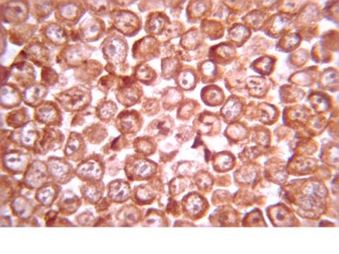
Immunohistochemistry Analysis: A 1:500 dilution from a representative lot detected RIP3 in rat pancreas tissue.

Qualità

Evaluated by Western Blot in human pancreas tissue lysate.

Western Blot Analysis: A 1:1,000 dilution of this antibody detected RIP3 in 10 µg of human pancreas tissue lysate.

Descrizione del bersaglio

~56 kDa (isoform 1) and ~27 kDa (isoform 2) observed.
Isoform 3 (~25 kDa) may be observed in some lysates. An uncharacterized band may be observed at ~16 kDa in some lysates.

Stato fisico

Format: Purified
Protein G Purified
Purified mouse monoclonal IgG2aκ in buffer containing 0.1 M Tris-Glycine (pH 7.4), 150 mM NaCl with 0.05% sodium azide.

Stoccaggio e stabilità

Stable for 1 year at 2-8°C from date of receipt.

Risultati analitici

Control
Human pancreas tissue lysate

Esclusione di responsabilità

Unless otherwise stated in our catalog or other company documentation accompanying the product(s), our products are intended for research use only and are not to be used for any other purpose, which includes but is not limited to, unauthorized commercial uses, in vitro diagnostic uses, ex vivo or in vivo therapeutic uses or any type of consumption or application to humans or animals.

Non trovi il prodotto giusto?  

Prova il nostro Motore di ricerca dei prodotti.

Codice della classe di stoccaggio

12 - Non Combustible Liquids

Classe di pericolosità dell'acqua (WGK)

WGK 1

Punto d’infiammabilità (°F)

Not applicable

Punto d’infiammabilità (°C)

Not applicable


Certificati d'analisi (COA)

Cerca il Certificati d'analisi (COA) digitando il numero di lotto/batch corrispondente. I numeri di lotto o di batch sono stampati sull'etichetta dei prodotti dopo la parola ‘Lotto’ o ‘Batch’.

Possiedi già questo prodotto?

I documenti relativi ai prodotti acquistati recentemente sono disponibili nell’Archivio dei documenti.

Visita l’Archivio dei documenti

Il team dei nostri ricercatori vanta grande esperienza in tutte le aree della ricerca quali Life Science, scienza dei materiali, sintesi chimica, cromatografia, discipline analitiche, ecc..

Contatta l'Assistenza Tecnica.