MAB4234
Anti-Wilms′ Tumor Antibody, NT, clone 6F-H2
clone 6F-H2, Chemicon®, from mouse
Sinonimo/i:
WT1
About This Item
Prodotti consigliati
Origine biologica
mouse
Livello qualitativo
Forma dell’anticorpo
purified immunoglobulin
Tipo di anticorpo
primary antibodies
Clone
6F-H2, monoclonal
Reattività contro le specie
human, mouse
Produttore/marchio commerciale
Chemicon®
tecniche
immunocytochemistry: suitable
immunofluorescence: suitable
immunohistochemistry: suitable (paraffin)
immunoprecipitation (IP): suitable
western blot: suitable
Isotipo
IgG1κ
N° accesso NCBI
N° accesso UniProt
Condizioni di spedizione
wet ice
modifica post-traduzionali bersaglio
unmodified
Informazioni sul gene
human ... WT1(7490)
Descrizione generale
Specificità
Immunogeno
Applicazioni
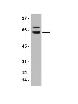
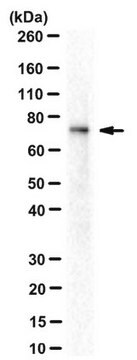
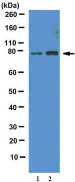
Western Blotting Analysis: A representative lot detected Wilms tumor protein WT1 in the CRE-binding protein/CBP immunoprecipitate obtained from the lysate of a T-SV40 immortalized human glomerular epithelial cell (HGEC) line (Drossopoulou, G.I., et al. (2009). Am. J. Physiol. Renal Physiol. 297(3):F594-F603).
Western Blotting Analysis: A representative lot detected Wilms tumor protein WT1 in lysates from mouse E15.5 embryonic kidney and human melanoma cell lines A375, SK-MEL-28, and WM-266-4 (Wagner, N., et al. (2008). Pflugers Arch. Eur. J. Physiol. 455(5):839-847).
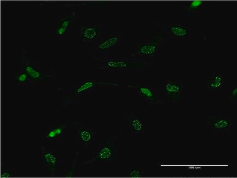
Immunocytochemistry Analysis: A representative lot immunostained the nucleus of methanol-fixed human melanoma A375 cells by fluorescent immunocytochemistry (Wagner, N., et al. (2008). Pflugers Arch. Eur. J. Physiol. 455(5):839-847).
Immunofluorescence Analysis: A representative lot immunostained the PCNA-positive nuclei of proliferating cells in formalin-fixed, paraffin-embedded human melanoma tissue sections by fluorescent immunohistochemistry (Wagner, N., et al. (2008). Pflugers Arch. Eur. J. Physiol. 455(5):839-847).
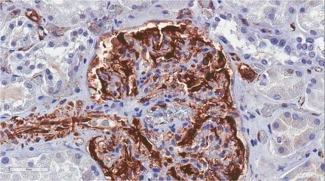
Immunohistochemistry Analysis: A representative lot immunostained glomeruli in formalin-fixed, paraffin-embedded normal human kidney and Wilms′ tumor sections (Wagner, N., et al. (2008). Pediatr. Nephrol. 23(9):1445-1453).
Immunohistochemistry Analysis: A representative lot detected vascular WT1 expression in 95% of 113 paraffin-embedded tumour tissues of various types. In most cases, nuclear WT1 staining of endothelial cells was seen (Wagner, N., et al. (2008). Oncogene. 27(26):3662-3672).
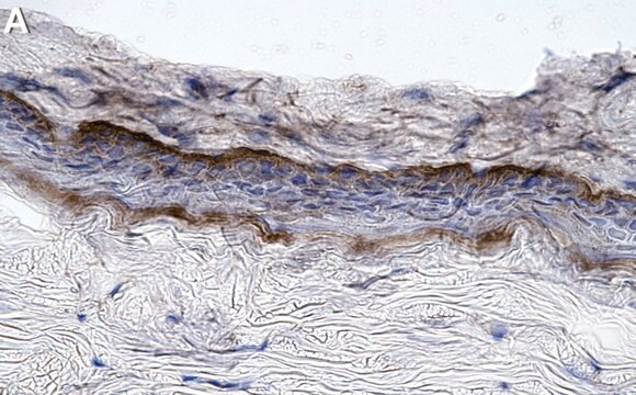
Immunohistochemistry Analysis: A representative lot immunostained the nucleus of perifollicular fibroblasts at the hair follicle in formalin-fixed, paraffin-embedded normal human skin sections. Most common melanocytic nevi do not express WT1, whereas Spitz nevi and dysplastic nevi show cytoplasmic WT1 staining. (Wagner, N., et al. (2008). Pflugers Arch. Eur. J. Physiol. 455(5):839-847).
Epigenetics & Nuclear Function
Transcription Factors
Descrizione del bersaglio
Stato fisico
Stoccaggio e stabilità
Altre note
Note legali
Esclusione di responsabilità
Non trovi il prodotto giusto?
Prova il nostro Motore di ricerca dei prodotti.
Raccomandato
Codice della classe di stoccaggio
10 - Combustible liquids
Classe di pericolosità dell'acqua (WGK)
WGK 2
Punto d’infiammabilità (°F)
Not applicable
Punto d’infiammabilità (°C)
Not applicable
Certificati d'analisi (COA)
Cerca il Certificati d'analisi (COA) digitando il numero di lotto/batch corrispondente. I numeri di lotto o di batch sono stampati sull'etichetta dei prodotti dopo la parola ‘Lotto’ o ‘Batch’.
Possiedi già questo prodotto?
I documenti relativi ai prodotti acquistati recentemente sono disponibili nell’Archivio dei documenti.
Il team dei nostri ricercatori vanta grande esperienza in tutte le aree della ricerca quali Life Science, scienza dei materiali, sintesi chimica, cromatografia, discipline analitiche, ecc..
Contatta l'Assistenza Tecnica.