Pular para o conteúdo
Merck
Todas as fotos(1)

Documentos Principais

MABC1092

Sigma-Aldrich

Anti-ASPP2 (TP53BP2) Antibody, clone DX54.10

clone DX54.10, from mouse

Sinônimo(s):

Apoptosis-stimulating of p53 protein 2, Bcl2-binding protein, Bbp, Renal carcinoma antigen NY-REN-51, Tumor suppressor p53-binding protein 2, 53BP2, p53-binding protein 2, p53BP2

Faça loginpara ver os preços organizacionais e de contrato


About This Item

Código UNSPSC:
12352203
eCl@ss:
32160702

fonte biológica

mouse

Nível de qualidade

forma do anticorpo

purified antibody

tipo de produto de anticorpo

primary antibodies

clone

DX54.10, monoclonal

reatividade de espécies

human

embalagem

antibody small pack of 25 μL

técnica(s)

immunocytochemistry: suitable
immunohistochemistry: suitable (paraffin)
immunoprecipitation (IP): suitable
western blot: suitable

Isotipo

IgG1κ

nº de adesão UniProt

Condições de expedição

ambient

modificação pós-traducional do alvo

unmodified

Informações sobre genes

human ... TP53BP2(7159)

Categorias relacionadas

Descrição geral

Apoptosis-stimulating of p53 protein 2 (UniProt: Q13625; also known as Bcl2-binding protein, Bbp, Renal carcinoma antigen NY-REN-51, Tumor suppressor p53-binding protein 2, 53BP2, p53-binding protein 2, p53BP2) is encoded by the TP53BP2 (also known as ASPP2, BBP) gene (Gene ID: 7159) in human. 53BP2 plays a central role in regulation of apoptosis and cell growth. It is widely expressed and its expression is observed in spleen, thymus, prostate, testis, ovary, small intestine, colon and peripheral blood leukocyte 53BP2 is induced following DNA damage by UV radiation. It is down-regulated by wild-type p53/TP53, but not by mutant p53/TP53. It regulates p53/TP53 action by enhancing the DNA binding and transactivation function of TP53 on the promoters of pro-apoptotic genes in vivo. 53BP2 is shown to inhibit the ability of APPBP1 to conjugate NEDD8 to CUL1, and thereby decreases APPBP1 ability to induce apoptosis. 53BP2 contains four ankyrin repeats and one SH3 domain are required for a specific interactions with TP53. The 53BP2 binding site on the p53 core domain consists of evolutionarily conserved regions that are frequently mutated in human cancers. Lower level of 53BP2 expression is observed in breast carcinomas expressing a wild-type TP53 protein.

Especificidade

Clone DX54.10 detects Apotosis -stimulating of p53 protein 2 (TP53BP2) in human cells. It targets an epitope with in 438 amino acids from the C-terminal half.

Imunogênio

Epitope: C-terminus
GST-tagged recombinant fragment corresponding to 438 amino acids from the C-terminal half of human Apoptosis-stimulating of p53 protein 2 (TP53BP2).

Aplicação

Anti-ASPP2 (TP53BP2), clone DX54.10, Cat. No. MABC1092, is a mouse monoclonal antibody that detects Apoptosis-stimulating of p53 protein 2 (ASPP2) and has been tested for use in Immunocytochemistry, Immunohistochemistry (Paraffin), Immunoprecipitation, and Western Blotting.
Immunoprecipitation Analysis: A representative lot detected ASPP2 (TP53BP2) in Immunoprecipitation applications (Samuels-Lev, Y., et. al. (2001). Mol Cell. 8(4):781-94).

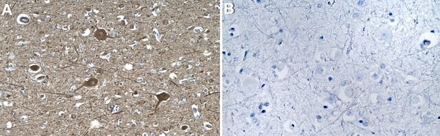
Immunohistochemistry Analysis: A representative lot detected ASPP2 (TP53BP2) in Immunohistochemistry applications (Wang, Y., et. al. (2014). Nat Cell Biol. 16(11):1092-104).

Immunocytochemistry Analysis: A representative lot detected ASPP2 (TP53BP2) in Immunocytohemistry applications (Wang, Y., et. al. (2013). Cell Death Differ. 20(4):525-34; Wang, Y., et. al. (2014). Nat Cell Biol. 16(11):1092-104).

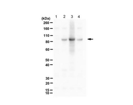
Western Blotting Analysis: A representative lot detected ASPP2 (TP53BP2) in Western Blotting applications (Wang, Y., et. al. (2013). Cell Death Differ. 20(4):525-34; Samuels-Lev, Y., et. al. (2001). Mol Cell. 8(4):781-94).
Research Category
Apoptosis & Cancer

Qualidade

Evaluated by Western Blotting in U2OS-iASPP and U2OS RISC-free cell lysates.

Western Blotting Analysis: A 1:125 dilution of this antibody detected ASPP2 in lysates of U2OS cells transfected with siRNA to knock down iASPP and in RISC-free U2OS cells.

Descrição-alvo

~170 kDa observed; 125.62 kDa calculated. Uncharacterized bands may be observed in some lysate(s).

forma física

Format: Purified
Protein G purified
Purified mouse monoclonal antibody IgG1 in buffer containing 0.1 M Tris-Glycine (pH 7.4), 150 mM NaCl with 0.05% sodium azide.

Armazenamento e estabilidade

Stable for 1 year at 2-8°C from date of receipt.

Outras notas

Concentration: Please refer to lot specific datasheet.

Exoneração de responsabilidade

Unless otherwise stated in our catalog or other company documentation accompanying the product(s), our products are intended for research use only and are not to be used for any other purpose, which includes but is not limited to, unauthorized commercial uses, in vitro diagnostic uses, ex vivo or in vivo therapeutic uses or any type of consumption or application to humans or animals.

Não está encontrando o produto certo?  

Experimente o nosso Ferramenta de seleção de produtos.

Código de classe de armazenamento

12 - Non Combustible Liquids

Classe de risco de água (WGK)

WGK 1

Ponto de fulgor (°F)

Not applicable

Ponto de fulgor (°C)

Not applicable


Certificados de análise (COA)

Busque Certificados de análise (COA) digitando o Número do Lote do produto. Os números de lote e remessa podem ser encontrados no rótulo de um produto após a palavra “Lot” ou “Batch”.

Já possui este produto?

Encontre a documentação dos produtos que você adquiriu recentemente na biblioteca de documentos.

Visite a Biblioteca de Documentos

Nossa equipe de cientistas tem experiência em todas as áreas de pesquisa, incluindo Life Sciences, ciência de materiais, síntese química, cromatografia, química analítica e muitas outras.

Entre em contato com a assistência técnica