Pular para o conteúdo
Merck
Todas as fotos(5)

Documentos Principais

MAB5356

Sigma-Aldrich

Anti-Rhodopsin Antibody, CT, last 9 amino acids, clone Rho 1D4

clone Rho 1D4, Chemicon®, from mouse

Sinônimo(s):

Anti-CSNBAD1, Anti-OPN2, Anti-RP4

Faça loginpara ver os preços organizacionais e de contrato


About This Item

Código UNSPSC:
12352203
eCl@ss:
32160702
NACRES:
NA.41

fonte biológica

mouse

Nível de qualidade

forma do anticorpo

purified immunoglobulin

tipo de produto de anticorpo

primary antibodies

clone

Rho 1D4, monoclonal

reatividade de espécies

vertebrates

fabricante/nome comercial

Chemicon®

técnica(s)

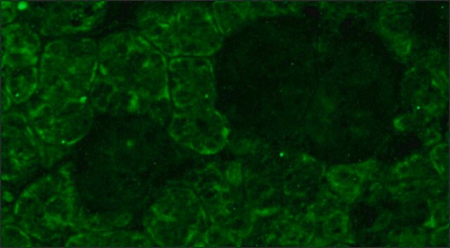
immunocytochemistry: suitable
immunohistochemistry: suitable
immunoprecipitation (IP): suitable
western blot: suitable

Isotipo

IgG1

nº de adesão NCBI

nº de adesão UniProt

Condições de expedição

wet ice

modificação pós-traducional do alvo

unmodified

Informações sobre genes

human ... RHO(6010)

Especificidade

Recognizes Rhodopsin. Reacts with both native and recombinant protein. No known reactivity to other proteins.

Imunogênio

Bovine rhodopsin
Epitope: C-terminus, last 9 amino acids

Aplicação

Anti-Rhodopsin Antibody, C-terminus, last 9 amino acids, clone Rho 1D4 detects level of Rhodopsin & has been published & validated for use in IC, IH, IP & WB.
Research Category
Neuroscience
Research Sub Category
Sensory & PNS
Western blot: 1:100-1:500 using ECL. Suggested dilution buffer is PBS and blocking buffer is PBS containing 5% milk. Recognizes 36 kDa monomers, dimmers and trimers depending on sample preparation. Boiling induces the aggregation of rhodopsin.

Western blot of isolated bovine rod outer segment labeled for rhodopsin (~85% of rod outer segment protein) with the rho 1D4 antibody. Amount of rod outer segments applied to the SDS gel: (a) 2.5 mg; (b) 0.63 mg; (c) 0.16 mg; (d) 0.04 mg. At higher protein quantities, dimers, trimers and tetramers of rhodopsin can be observed along with the monomer.

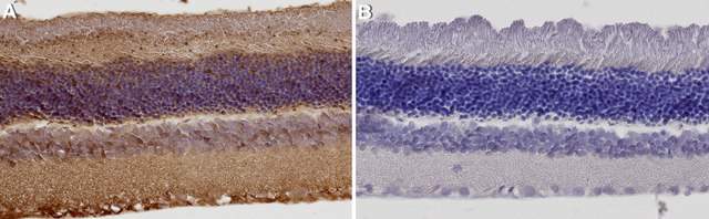
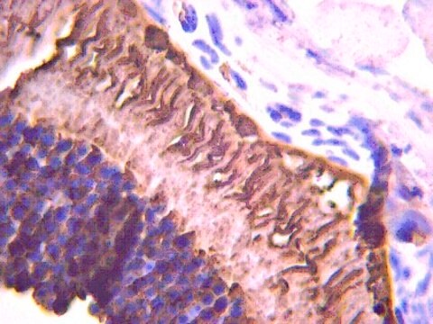
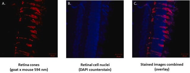
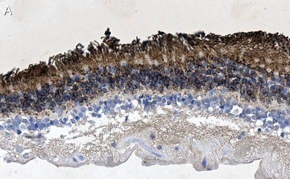
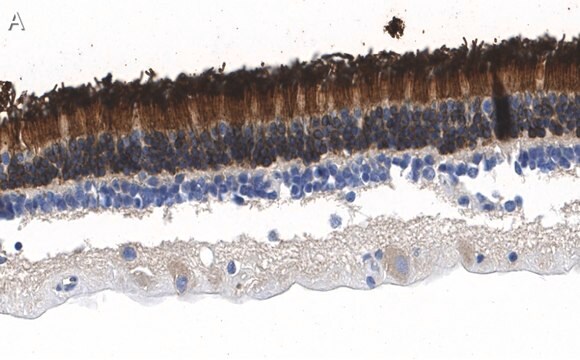
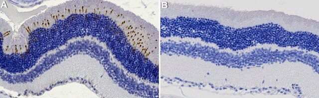
Immunohisto/cytochemistry: 1:100-1:500 on paraformaldehyde and glutaraldehyde fixed frozen tissue sections. Preferred fixation is paraformaldehyde for 4 hours at 2-8°C. Suggested permeabilization method is 0.2% Triton X-100.

Immunoprecipitation

Optimal working dilutions must be determined by the end user.

Descrição-alvo

48 kDa

forma física

Format: Purified
Protein A Purified mouse immunoglobulin in 20 mM sodium phosphate, 250 mM NaCl, pH. 7.6, with 0.1% sodium azide as a preservative.
Protein A purified

Armazenamento e estabilidade

Maintain for 1 year at 2–8°C from date of shipment. Aliquot to avoid repeated freezing and thawing. For maximum recovery of product, centrifuge the original vial after thawing and prior to removing the cap.

Nota de análise

Control
Eye

Outras notas

Concentration: Please refer to the Certificate of Analysis for the lot-specific concentration.

Informações legais

CHEMICON is a registered trademark of Merck KGaA, Darmstadt, Germany

Exoneração de responsabilidade

Unless otherwise stated in our catalog or other company documentation accompanying the product(s), our products are intended for research use only and are not to be used for any other purpose, which includes but is not limited to, unauthorized commercial uses, in vitro diagnostic uses, ex vivo or in vivo therapeutic uses or any type of consumption or application to humans or animals.

Não está encontrando o produto certo?  

Experimente o nosso Ferramenta de seleção de produtos.

Código de classe de armazenamento

10 - Combustible liquids

Classe de risco de água (WGK)

WGK 2

Ponto de fulgor (°F)

Not applicable

Ponto de fulgor (°C)

Not applicable


Certificados de análise (COA)

Busque Certificados de análise (COA) digitando o Número do Lote do produto. Os números de lote e remessa podem ser encontrados no rótulo de um produto após a palavra “Lot” ou “Batch”.

Já possui este produto?

Encontre a documentação dos produtos que você adquiriu recentemente na biblioteca de documentos.

Visite a Biblioteca de Documentos

M Keith Wyatt et al.
PloS one, 8(6), e68088-e68088 (2013-07-11)
Age-related macular degeneration (AMD) is a major cause of vision loss. It is associated with development of characteristic plaque-like deposits (soft drusen) in Bruch's membrane basal to the retinal pigment epithelium (RPE). A sequence variant (Y402H) in short consensus repeat
A novel experimental mouse model of retinal detachment: complete functional and histologic recovery of the retina.
Zeng, R; Zhang, Y; Shi, F; Kong, F
Investigative Ophthalmology & Visual Science null
S Nagar et al.
Neuroscience, 160(2), 517-529 (2009-03-11)
Photoreceptor degeneration is followed by significant morphological changes in the second-order retinal neurons in humans and in several genetic animal models. However, it is not clear whether similar changes occur when photoreceptor degeneration is induced nongenetically, raising the question whether
Genetic dissection of TAM receptor-ligand interaction in retinal pigment epithelial cell phagocytosis.
Burstyn-Cohen, T; Lew, ED; Traves, PG; Burrola, PG; Hash, JC; Lemke, G
Neuron null
Jun Yin et al.
Investigative ophthalmology & visual science, 53(8), 4943-4951 (2012-06-30)
In experimental eye research, zebrafish has become a powerful model for human retina disorders. The purpose of the present study is the characterization of antibodies commonly employed in zebrafish models for rod photoreceptor degeneration. The 1D4 monoclonal antibody, developed against

Nossa equipe de cientistas tem experiência em todas as áreas de pesquisa, incluindo Life Sciences, ciência de materiais, síntese química, cromatografia, química analítica e muitas outras.

Entre em contato com a assistência técnica