Pular para o conteúdo
Merck
Todas as fotos(3)

Key Documents

MAB5258

Sigma-Aldrich

Anti-Synaptophysin Antibody, clone SY38

clone SY38, Chemicon®, from mouse

Sinônimo(s):

synaptophysin, Synaptophysin

Faça loginpara ver os preços organizacionais e de contrato


About This Item

Código UNSPSC:
12352203
eCl@ss:
32160702
NACRES:
NA.41

fonte biológica

mouse

Nível de qualidade

forma do anticorpo

purified immunoglobulin

tipo de produto de anticorpo

primary antibodies

clone

SY38, monoclonal

reatividade de espécies

bovine, rat, avian, fish, mouse, human

fabricante/nome comercial

Chemicon®

técnica(s)

flow cytometry: suitable
immunocytochemistry: suitable
immunohistochemistry: suitable
western blot: suitable

Isotipo

IgG1κ

nº de adesão NCBI

nº de adesão UniProt

Condições de expedição

wet ice

modificação pós-traducional do alvo

unmodified

Informações sobre genes

human ... SYP(6855)

Descrição geral

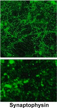
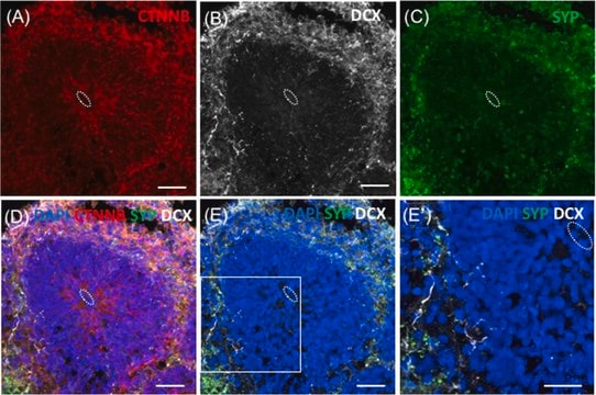
Synaptophysin is a 38 kDa glycoprotein present in the membrane of neuronal presynaptic vesicles in brain, spinal cord, retina, vesicles of adrenal medulla, neuromuscular junctions, and endocrine cells. It is also expressed by neuroendocrine cells throughout the body, both normal and neoplastic. Synaptophysin is a useful marker for the identification of normal neuroendocrine cells and neuroendocrine neoplasms. This antibody reacts with neuroendocrine cells of human adrenal medulla, carotid body, skin, pituitary gland, thyroid, lung, pancreas, and gastointestinal mucosa. It also reacts with a wide spectrum of neuroendocrine neoplasms of neural type including neuroblastomas, ganglioneuroblastomas, ganglioneuromas, pheochromocytomas, chromaffin, and non chromaffin paragangliomas.

Especificidade

The antibody reacts with presynaptic vesicles of cerebral and spinal neurons, of neuromuscular endplates and with the retina of man, cow, rat and mouse. The antibody also reacts with vesicles of adrenal medulla and islet cells, and additionally allows specific staining of neuronal, adrenal and neuroepithelial tumors, such as pheochromocytoma, paraganglioma, islet cell tumors (incl. Insulinoma), medullary thyroid carcinoma and diverse pulmonary and gastro-intestinal carcinoids. The antibody also stains neurosecretory vesicles of certain culture cells, e.g. of the rat cell line PC-12.

Imunogênio

Vesicular fraction of bovine brain. The SY38 epitope is to a pentapeptide repeat structure in the carboxy-terminal cytoplasmic tail of synaptophysin (Knaus & Betz 1990).

Aplicação

Detect Synaptophysin using this Anti-Synaptophysin Antibody, clone SY38 validated for use in FC, IC, IH & WB.
Flow Cytometry (FACS):
A previous lot of this antibody was used in Flow Cytometry.

Western Blot:
(Provoda, 2000) Reacts with a 38 kDa transmembrane glycoprotein

Immunohistochemistry:
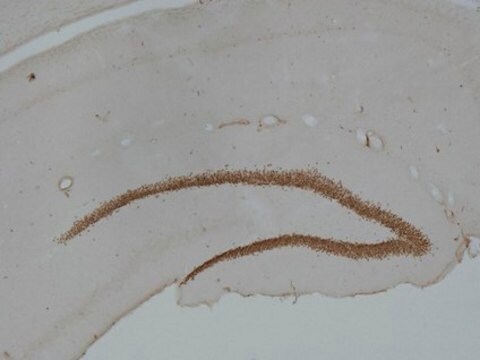
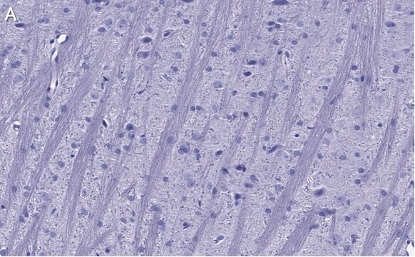
For relatively cytoplasm-rich neuroendocrinic tumors a final concentration of 1 µg/mL is recommended. Concerning cytoplasm deficient tumors a concentration of 2 µg/mL should be used. Ideal frozen sections (4-5 mm) are obtained from shock-frozen tissue samples. The frozen sections are air-dried and then fixed with acetone for 5-10 min at -20°C. Excess acetone is allowed to evaporate at room temperature. Material fixed in alcohol or formalin and embedded in paraffin can also be used.
It is advantageous to block unspecific binding sites by overlaying the sections with fetal calf serum for 20-30 min at room temperature. Excess of fetal calf serum is removed by decanting before application of the anti-body solution.
Cytocentrifuge preparations of single cells or cell smears are also fixed in acetone. These preparations should but should be followed directly by antibody treatment.

Optimal working dilutions must be determined by end user.
Research Category
Neuroscience
Research Sub Category
Synapse & Synaptic Biology

Qualidade

Routinely evaluated by Western Blot on mouse brain lysates.

Western Blot Analysis:
1:1000 dilution of this lot detected synaptophysin on 10 μg of mouse brain lysates.

Descrição-alvo

38 kDa

Ligação

Replaces: 04-1019

forma física

Format: Purified
Protein A purified
Purified mouse monoclonal IgG1κ in buffer containing 20 mM sodium phosphate buffer, 0.25 M NaCl, containing 0.1% sodium azide.

Armazenamento e estabilidade

Stable for 6 months at 2-8ºC from date of receipt. DO NOT FREEZE.

Nota de análise

Control
Positive Control: Pancreas tissue
Negative Control: Normal mouse serum
Rat hippocampus tissue, mouse brain lysate.

Outras notas

Concentration: Please refer to the Certificate of Analysis for the lot-specific concentration.

Informações legais

CHEMICON is a registered trademark of Merck KGaA, Darmstadt, Germany

Exoneração de responsabilidade

Unless otherwise stated in our catalog or other company documentation accompanying the product(s), our products are intended for research use only and are not to be used for any other purpose, which includes but is not limited to, unauthorized commercial uses, in vitro diagnostic uses, ex vivo or in vivo therapeutic uses or any type of consumption or application to humans or animals.

Não está encontrando o produto certo?  

Experimente o nosso Ferramenta de seleção de produtos.

Código de classe de armazenamento

10 - Combustible liquids

Classe de risco de água (WGK)

WGK 2

Ponto de fulgor (°F)

Not applicable

Ponto de fulgor (°C)

Not applicable


Certificados de análise (COA)

Busque Certificados de análise (COA) digitando o Número do Lote do produto. Os números de lote e remessa podem ser encontrados no rótulo de um produto após a palavra “Lot” ou “Batch”.

Já possui este produto?

Encontre a documentação dos produtos que você adquiriu recentemente na biblioteca de documentos.

Visite a Biblioteca de Documentos

Claudia Fuchs et al.
Neurobiology of disease, 70, 53-68 (2014-06-22)
Mutations in the X-linked cyclin-dependent kinase-like 5 (CDKL5) gene have been identified in a neurodevelopmental disorder characterized by early-onset intractable seizures, severe developmental delay, intellectual disability, and Rett's syndrome-like features. Since the physiological functions of CDKL5 still need to be
Sarah L Roche et al.
Investigative ophthalmology & visual science, 60(8), 3221-3235 (2019-07-25)
Retinitis pigmentosa (RP) refers to a group of inherited blinding retinal diseases, whereby the death of mutated rod photoreceptors is followed closely by the death of cone photoreceptors. Cone cell death can be hugely debilitating as color/daytime vision becomes impaired.
Paul T Tarr et al.
Journal of lipid research, 49(1), 169-182 (2007-10-06)
Here, we describe the initial characterization of Abcg4(-/-) mice and identify overlapping functions of ABCG4 and ABCG1 in the brain. Histological examination of tissues from Abcg4(+/-)/nlsLacZ and Abcg1(+/-)/nlsLacZ mice demonstrates that coexpression of Abcg4 and Abcg1 is restricted to neurons
Gloria Salazar et al.
Molecular biology of the cell, 16(8), 3692-3704 (2005-06-10)
A membrane fraction enriched in vesicles containing the adaptor protein (AP) -3 cargo zinc transporter 3 was generated from PC12 cells and was used to identify new components of these organelles by mass spectrometry. Proteins prominently represented in the fraction
Hengchang Li et al.
Frontiers in neuroscience, 13, 706-706 (2019-07-30)
Children, after multiple exposures to general anesthesia, appear to be at an increased risk of developing learning disabilities. Almost all general anesthetics-including sevoflurane, which is commonly used for children-are potentially neurotoxic to the developing brain. Anesthesia exposure during development might

Nossa equipe de cientistas tem experiência em todas as áreas de pesquisa, incluindo Life Sciences, ciência de materiais, síntese química, cromatografia, química analítica e muitas outras.

Entre em contato com a assistência técnica