Accéder au contenu
Merck
Toutes les photos(1)

Principaux documents

281M-9

Sigma-Aldrich

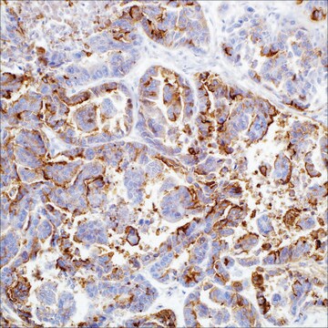
MART-1 (Melan A) (M2-7C10) Mouse Monoclonal Antibody

Se connecterpour consulter vos tarifs contractuels et ceux de votre entreprise/organisme


About This Item

Code UNSPSC :
12352203
Nomenclature NACRES :
NA.41
Clone:
M2-7C10, monoclonal
application:
IHC (p)
Espèces réactives:
human
Technique(s):
immunohistochemistry (formalin-fixed, paraffin-embedded sections): 1:100-1:500
citations:
8

Source biologique

mouse

Niveau de qualité

100
500

Conjugué

unconjugated

Forme d'anticorps

culture supernatant

Type de produit anticorps

primary antibodies

Clone

M2-7C10, monoclonal

Description

For In Vitro Diagnostic Use in Select Regions (See Chart)

Forme

buffered aqueous solution

Espèces réactives

human

Conditionnement

vial of 0.1 mL concentrate (281M-94)
vial of 0.5 mL concentrate (281M-95)
bottle of 1.0 mL predilute (281M-97)
vial of 1.0 mL concentrate (281M-96)
bottle of 7.0 mL predilute (281M-98)

Fabricant/nom de marque

Cell Marque

Technique(s)

immunohistochemistry (formalin-fixed, paraffin-embedded sections): 1:100-1:500

Isotype

IgG2bκ

Contrôle

melanoma

Conditions d'expédition

wet ice

Température de stockage

2-8°C

Visualisation

cytoplasmic

Informations sur le gène

human ... MLANA(2315)

Description générale

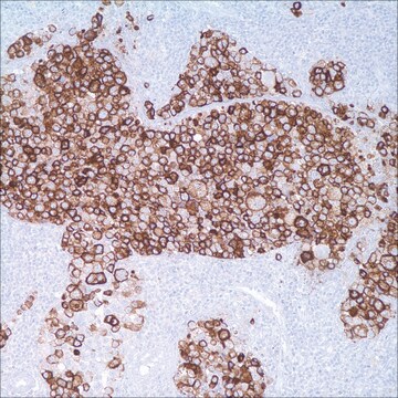
MART-1 (also known as Melan A) is a melanocyte differentiation antigen. It is present in melanocytes of normal skin, retina, nevi, and in more than 85% of melanomas. This antibody is very useful in establishing the diagnosis of metastatic melanomas.

Liaison

MART-1 Positive Control Slides, Product No. 281S, are available for immunohistochemistry (formalin-fixed, paraffin-embedded sections).

Forme physique

Solution in Tris Buffer, pH 7.3-7.7, with 1% BSA and <0.1% Sodium Azide

Notes préparatoires

Download the IFU specific to your product lot and formatNote: This requires a keycode which can be found on your packaging or product label.

Autres remarques

For Technical Service please contact: 800-665-7284 or email: service@cellmarque.com

Informations légales

Cell Marque is a trademark of Merck KGaA, Darmstadt, Germany

Vous ne trouvez pas le bon produit ?  

Essayez notre Outil de sélection de produits.


Faites votre choix parmi les versions les plus récentes :

Certificats d'analyse (COA)

Lot/Batch Number

Vous ne trouvez pas la bonne version ?

Si vous avez besoin d'une version particulière, vous pouvez rechercher un certificat spécifique par le numéro de lot.

Déjà en possession de ce produit ?

Retrouvez la documentation relative aux produits que vous avez récemment achetés dans la Bibliothèque de documents.

Consulter la Bibliothèque de documents

Z Orosz
Histopathology, 34(6), 517-525 (1999-06-26)
The purpose of this study was to test different malignant non-melanocytic tumours with the commercially available antibody Melan-A to examine its diagnostic specificity and to compare the S100, Melan-A and HMB-45 reactivity in various melanocytic lesions. Seventy-three benign and malignant
R Bergman et al.
Journal of the American Academy of Dermatology, 42(3), 496-500 (2000-02-25)
The histopathologic differential diagnosis of Spitz nevus (SN) from malignant melanoma (MM) may be difficult. We attempted to elucidate the pattern of expression of a newly recognized melanocyte-specific melanosomal protein MART-1 in routinely processed specimens of SNs, MMs, and ordinary
P A Fetsch et al.
Cancer, 87(1), 37-42 (1999-03-30)
HMB-45, an antibody directed against a premelanosome glycoprotein, has thus far been considered the most specific antibody for the immunocytochemical substantiation of the diagnosis of malignant melanoma (MM). A recently described antigen, MART-1, is a transmembrane protein that is present
S Mocellin et al.
Journal of immunotherapy (Hagerstown, Md. : 1997), 24(6), 447-458 (2002-01-05)
Assessment of antigen expression by solid tumors has relied predominantly on immunohistochemistry, flow cytometry, and more recently quantitative real-time polymerase chain reaction. However, all these techniques present intrinsic limits. The laser scanning cytometer, by combining the properties of light and
T Kageshita et al.
Journal of immunotherapy (Hagerstown, Md. : 1997), 20(6), 460-465 (1997-12-31)
Twenty-eight primary and 29 metastatic melanoma lesions and 18 pigmented nevi lesions were analyzed by using the immunoperoxidase reaction with anti-MART-1 and anti-gp100 monoclonal antibodies (mAbs). The MART-1 was expressed in 28, 29, and 18, and gp100 was expressed in

Articles

Immunohistochemistry (IHC) techniques and applications have greatly improved, dermatopathology is still largely based on H&E stained slides.This paper outlines ways in which IHC antibodies can be utilized for dermatopathology.

Notre équipe de scientifiques dispose d'une expérience dans tous les secteurs de la recherche, notamment en sciences de la vie, science des matériaux, synthèse chimique, chromatographie, analyse et dans de nombreux autres domaines..

Contacter notre Service technique