S7100
ApopTag Peroxidase In-situ-Apoptosenachweis-Kit
The ApopTag Peroxidase In Situ Apoptosis Detection Kit detects apoptotic cells in situ by labeling & detecting DNA strand breaks by the TUNEL method.
Synonym(e):
ApopTag Kit, In Situ Apoptosis Detection Kit
About This Item
Empfohlene Produkte
Qualitätsniveau
Hersteller/Markenname
ApopTag
Chemicon®
Nachweisverfahren
colorimetric
Versandbedingung
dry ice
Allgemeine Beschreibung
Of all the aspects of apoptosis, the defining characteristic is a complete change in cellular morphology. As observed by electron microscopy, the cell undergoes shrinkage, chromatin margination, membrane blebbing, nuclear condensation and then segmentation, and division into apoptotic bodies which may be phagocytosed (11, 19, 24). The characteristic apoptotic bodies are short-lived and minute, and can resemble other cellular constituents when viewed by brightfield microscopy. DNA fragmentation in apoptotic cells is followed by cell death and removal from the tissue, usually within several hours (7). A rate of tissue regression as rapid as 25% per day can result from apparent apoptosis in only 2-3% of the cells at any one time (6). Thus, the quantitative measurement of an apoptotic index by morphology alone can be difficult.
DNA fragmentation is usually associated with ultrastructural changes in cellular morphology in apoptosis (26, 38). In a number of well-researched model systems, large fragments of 300 kb and 50 kb are first produced by endonucleolytic degradation of higher-order chromatin structural organization. These large DNA fragments are visible on pulsed-field electrophoresis gels (5, 43, 44). In most models, the activation of Ca2+- and Mg2+-dependent endonuclease activity further shortens the fragments by cleaving the DNA at linker sites between nucleosomes (3). The ultimate DNA fragments are multimers of about 180 bp nucleosomal units. These multimers appear as the familiar "DNA ladder" seen on standard agarose electrophoresis gels of DNA extracted from many kinds of apoptotic cells (e.g. 3, 7,13, 35, 44).
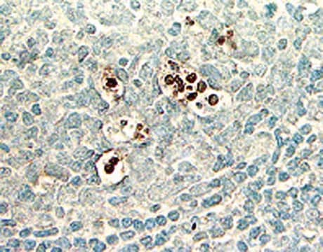
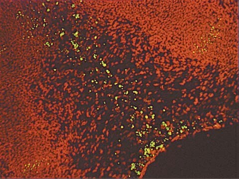
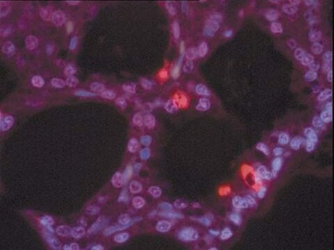
Another method for examining apoptosis via DNA fragmentation is by the TUNEL assay, (13) which is the basis of ApopTag technology. The DNA strand breaks are detected by enzymatically labeling the free 3′-OH termini with modified nucleotides. These new DNA ends that are generated upon DNA fragmentation are typically localized in morphologically identifiable nuclei and apoptotic bodies. In contrast, normal or proliferative nuclei, which have relatively insignificant numbers of DNA 3′-OH ends, usually do not stain with the kit. ApopTag Kits detect single-stranded (25) and double-stranded breaks associated with apoptosis. Drug-induced DNA damage is not identified by the TUNEL assay unless it is coupled to the apoptotic response (8). In addition, this technique can detect early-stage apoptosis in systems where chromatin condensation has begun and strand breaks are fewer, even before the nucleus undergoes major morphological changes (4, 8).
Apoptosis is distinct from accidental cell death (necrosis). Numerous morphological and biochemical differences that distinguish apoptotic from necrotic cell death are summarized in the following table (adapted with permission from reference 39).
Komponenten
Reaktionspuffer 2,0 ml -15 °C bis -25 °C
TdT-Enzym 0,64 ml -15 °C bis -25 °C
Stopp-/Waschpuffer 20 ml -15 °C bis -25 °C
Anti-Digoxigenin-Peroxidase* 3,0 ml 2 °C bis 8 °C
Kunststoff-Deckgläschen je 100 St. Raumtemperatur
Hinweis: DAB (Peroxidasesubstrat) muss separat erworben werden. Es ist nicht im Lieferumfang dieses Kits enthalten.
Anzahl der Produktmuster pro Kit: Es werden ausreichend viele Materialien zur Verfügung gestellt, um bei einer anleitungsgemäßen Verwendung 40 Gewebeproben von jeweils ca. 5 mm2 zu färben. Wenn die Kits für Proben auf Objektträgern verwendet werden, wird der Reaktionspuffer vor Einsatz anderer Reagenzien vollständig aufgebraucht.
Lagerung und Haltbarkeit
Vorsichtshinweise
1. Die folgenden Komponenten des Kits enthalten Kaliumcacodylat (Dimethylarsinsäure) als Puffer: Äquilibrierungspuffer (90416), Reaktionspuffer (90417) und TdT-Enzym (90418). Diese Komponenten sind bei Verschlucken gesundheitsschädlich; Kontakt mit Haut und Augen vermeiden (Handschuhe, Schutzbrille tragen) und Kontaktbereiche sofort abwaschen.
2. Antikörperkonjugate (90420) und Blockierlösungen (Nr. 10 und Nr. 13) enthalten 0,08 % Natriumazid als Konservierungsmittel.
3. TdT-Enzym (90418) enthält Glycerin und gefriert bei -20 °C nicht. Für eine maximale Haltbarkeitsdauer dieses Reagenz vor der Entnahme nicht auf Raumtemperatur erwärmen.
Rechtliche Hinweise
Signalwort
Danger
H-Sätze
Gefahreneinstufungen
Aquatic Chronic 2 - Carc. 1B - Skin Sens. 1 - STOT RE 2 Inhalation
Zielorgane
Respiratory Tract
Lagerklassenschlüssel
6.1C - Combustible acute toxic Cat.3 / toxic compounds or compounds which causing chronic effects
Analysenzertifikate (COA)
Suchen Sie nach Analysenzertifikate (COA), indem Sie die Lot-/Chargennummer des Produkts eingeben. Lot- und Chargennummern sind auf dem Produktetikett hinter den Wörtern ‘Lot’ oder ‘Batch’ (Lot oder Charge) zu finden.
Besitzen Sie dieses Produkt bereits?
In der Dokumentenbibliothek finden Sie die Dokumentation zu den Produkten, die Sie kürzlich erworben haben.
Kunden haben sich ebenfalls angesehen
Artikel
Cellular apoptosis assays to detect programmed cell death using Annexin V, Caspase and TUNEL DNA fragmentation assays.
Unser Team von Wissenschaftlern verfügt über Erfahrung in allen Forschungsbereichen einschließlich Life Science, Materialwissenschaften, chemischer Synthese, Chromatographie, Analytik und vielen mehr..
Setzen Sie sich mit dem technischen Dienst in Verbindung.