Urine Concentration with Amicon® Ultra Centrifugal Filters
Accurate measurement of specific proteins in urine is important for diagnosing and managing diseases. However, the concentration of urine proteins is often below the detection limit of clinical tests. Amicon® Ultra-4 mL 10 kDa MWCO devices can be used to concentrate urine samples prior to clinical laboratory analyses.
Method
Enrichment of free immunoglobulin light chains (Bence-Jones proteins) in urine samples using Amicon® Ultra-4 mL 10 kDa MWCO filters.
- Determine the total protein in a 24-hour urine specimen.
- Fill Amicon® Ultra-4mL device with 4 mL of urine.
- Centrifuge at 3400 x g for 30–45 minutes (until approximately 25–50 μL of concentrated sample is obtained). This represents a 160-fold increase in concentration of the original sample.
- Insert a pipette tip into the bottom of the filter unit and withdraw the concentrated sample.
- Perform agarose electrophoresis on the concentrate to quantitate light chains and identify other proteins. Determine the percentage of light chains with respect to the total number of components in the urine. Then multiply the percentage of light chains by the total 24-hour protein concentration (grams per 24-hour volume).
- Perform immunofixation electrophoresis on the concentrate to identify light chains.

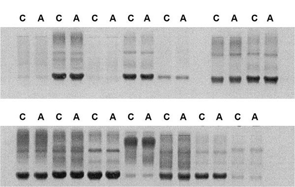
Results of electrophoretic resolution of free immunoglobulin light chains (Bence-Jones proteins) in urine samples concentrated using Centricon® (C) and Amicon® Ultra (A) centrifugal concentration devices. The dark bands at ~25 kDa in each lane represent these proteins.
Additional Notes
- Normal heterogeneous immunoglobulins may also be seen in urine concentrate with immunofixation electrophoresis. This “ladder effect” is comprised of microheterogeneous light chains. Bence-Jones proteins may be within this ladder. To verify the presence of Bence-Jones proteins requires additional analysis by two-dimensional electrophoresis.
- If there is excess antigen, dilution of the concentrate will be required until equilibrium is achieved between the antigen (Bence-Jones protein) and the antibody.
Related Products
- Amicon® Ultra devices can also be used to concentrate serum, plasma and cerebrospinal fluid for similar analyses. A concentration of approximately 20 mg/mL is required in order to detect free light chains from diseased patients by agarose electrophoresis. Detection by immunofixation electrophoresis is 10 times more sensitive than by agarose electrophoresis.
- Static concentrators (Minicon® devices) are also available for concentration of Bence-Jones protein in urine.
Acknowledgements
Research using Amicon® Ultra devices for urine concentration in this protocol was conducted by Mark Merchant, Ph.D. at Helena Laboratories, Beaumont, TX.
References
To continue reading please sign in or create an account.
Don't Have An Account?