MABN1786
Anti-Merlin (NF2) Antibody, clone 1C4
clone 1C4, from mouse
Sinónimos:
Merlin, Moesin-ezrin-radixin-like protein, Neurofibromin-2, Schwannomerlin, Schwannomin
About This Item
Productos recomendados
biological source
mouse
Quality Level
antibody form
purified antibody
antibody product type
primary antibodies
clone
1C4, monoclonal
species reactivity
rat, mouse, human
technique(s)
immunocytochemistry: suitable
immunoprecipitation (IP): suitable
western blot: suitable
isotype
IgG1κ
NCBI accession no.
UniProt accession no.
shipped in
ambient
target post-translational modification
unmodified
Gene Information
human ... NF2(4771)
General description
Specificity
Immunogen
Application
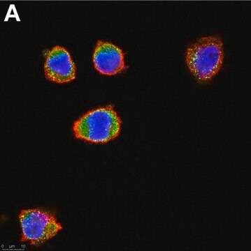
Immunocytochemistry Analysis: A representative lot detected merlin cellular distribution and HEI10 co-localization in a cell cycle-dependent manner by fluorescent immunocytochemistry staining of 3.5% paraformaldehyde-fixed U2OS human osteosarcoma cells (Grönholm, M., et al. (2006). Oncogene. 25(32):4389-4398).
Immunocytochemistry Analysis: A representative lot detected merlin cellular localization by fluorescent immunocytochemistry staining of 3.5% paraformaldehyde-fixed embryonic E16 rat neurons. Merlin was seen co-localized with RI to the cell body and extensions in a punctate pattern (Grönholm, M., et al. (2003). J. Biol. Chem. 278(42):41167-41172).
Immunocytochemistry Analysis: A representative lot detected merlin cellular localization by fluorescent immunocytochemistry staining of 4% paraformaldehyde-fixed, 0.1 NP-40-permeabilized human fibroblasts and primary meningioma cells. Merlin co-localized with F-actin at the leading and ruffling edges, but not at the stress fiber. No merlin co-localization with ezrin or moesin was observed (Gonzalez-Agosti, C., et al. (1996). Oncogene. 13(6):1239-1247).
Immunoprecipitation Analysis: A representative lot co-immunoprecipitated N-WASP with merlin from HEK293T cell lysate (Manchanda, N., et al. (2005). J. Biol. Chem. 280(13):12517-12522).
Western Blotting Analysis: A representative lot detected the presence of wild-type, but not L64P, merlin in the EGFR immunoprecipitates from Nf2-/- MEFs infected by adenovirus to express either wild-type or L64P merlin (Curto, M., et al. (2007). J. Cell Biol. 177(5):893-903).
Western Blotting Analysis: A representative lot detected a reduced merlin level in EGFR immunoprecipitate from mouse liver-derived epithelial cells (LDCs) upon shRNA-mediated NHE-RF1, but not NHE-RF2 knockdown. Merlin association with Ezrin was not affected by NHE-RF1 knockdown (Curto, M., et al. (2007). J. Cell Biol. 177(5):893-903).
Western Blotting Analysis: A representative lot detected a ~72 kDa merlin band in rat newborn fibroblast (RNF) and S-16 rat Schwann cell lysates, as well as a ~66 kDa merlin band in murine NIH/3T3 and human MRC-5 fetal fibroblast lystes without cross-reactivity toward the three ERM protein family members, ezrin, radixin, and moesin (Gonzalez-Agosti, C., et al. (1996). Oncogene. 13(6):1239-1247).
Neuroscience
Quality
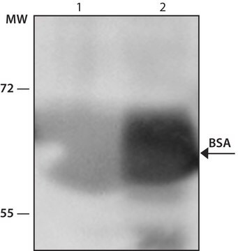
Western Blotting Analysis: 4 µg/mL of this antibody detected Merlin (NF2) in 10 µg of HEK293 cell lysate.
Target description
Physical form
Storage and Stability
Other Notes
Disclaimer
¿No encuentra el producto adecuado?
Pruebe nuestro Herramienta de selección de productos.
Storage Class
12 - Non Combustible Liquids
wgk_germany
WGK 1
flash_point_f
Not applicable
flash_point_c
Not applicable
Certificados de análisis (COA)
Busque Certificados de análisis (COA) introduciendo el número de lote del producto. Los números de lote se encuentran en la etiqueta del producto después de las palabras «Lot» o «Batch»
¿Ya tiene este producto?
Encuentre la documentación para los productos que ha comprado recientemente en la Biblioteca de documentos.
Nuestro equipo de científicos tiene experiencia en todas las áreas de investigación: Ciencias de la vida, Ciencia de los materiales, Síntesis química, Cromatografía, Analítica y muchas otras.
Póngase en contacto con el Servicio técnico


