ABN991
抗磷酸GLUT-1抗体(Ser226)
from rabbit, purified by affinity chromatography
别名:
Solute carrier family 2, facilitated glucose transporter member 1, Glucose transporter type 1, erythrocyte/brain, GLUT-1, HepG2 glucose transporter, Human T-cell leukemia virus I and II receptor, Receptor for HTLV-1 and HTLV-2, phospho GLUT-1 (Ser226)
About This Item
推荐产品
生物源
rabbit
品質等級
抗體表格
affinity isolated antibody
抗體產品種類
primary antibodies
無性繁殖
polyclonal
純化經由
affinity chromatography
物種活性
mouse, human
物種活性(以同源性預測)
horse (based on 100% sequence homology), canine (based on 100% sequence homology), bovine (based on 100% sequence homology), Xenopus (based on 100% sequence homology), rat (based on 100% sequence homology), rabbit (based on 100% sequence homology)
技術
immunocytochemistry: suitable
inhibition assay: suitable (peptide)
western blot: suitable
NCBI登錄號
UniProt登錄號
運輸包裝
wet ice
目標翻譯後修改
phosphorylation (pSer226 )
基因資訊
human ... SLC2A1(6513)
一般說明
免疫原
應用
蛋白质印迹分析:代表性批次的1:200稀释液(5 µg/ml)在来自Rat2成纤维细胞的裂解物中检测到TPA诱导的野生型GLUT-1磷酸化,但未检测到带有S226A突变的GLUT-1磷酸化,Rat2成纤维细胞通过逆转录病毒介导的转染表达相应结构(由Dr. Richard C. Wang, UT Southwestern Medical Center, Dallas, TX提供)。
蛋白质印迹分析:代表性批次的1:200稀释液(5 µg/ml)在VEGF处理后,在血清饥饿的人脐静脉内皮细胞(HUVEC)和人主动脉内皮细胞(HAEC)中检测到时间依赖性GLUT-1 Ser226磷酸化诱导(由Dr. Richard C. Wang, UT Southwestern Medical Center, Dallas, TX提供)。
蛋白质印迹分析:代表性批次仅在不存在PKC抑制剂Go 6983(目录号365251)的情况下,在TPA(目录号500582 &524400)处理后检测到人主动脉内皮细胞(HAEC)中PKC激活诱导的GLUT-1 Ser226磷酸化,在存在PKC抑制剂Go 6983的情况下未检测到(Lee, E.E., et al. (2015).Mol.Cell.58(5):845-853)。
蛋白质印迹分析:代表性批次在VEGF或血管紧张肽II处理后,在血清饥饿的人脐静脉内皮细胞(HUVEC)中检测到了时间依赖性GLUT-1 Ser226磷酸化诱导(Lee, E.E., et al. (2015).Mol.Cell.58(5):845-853)。
蛋白质印迹分析:代表性批次在PKC激活剂TPA(目录号500582&524400)处理(目录号365251)后在转染的大鼠2成纤维细胞中检测到外源表达的野生型GLUT-1或K526E突变体的可比性Ser226磷酸化诱导(Lee, E.E., et al. (2015).Mol.Cell.58(5):845-853)。
蛋白质印迹分析:代表性批次在血清饥饿HeLa、人原代心脏内皮细胞、EA.hy926人内皮细胞和bEnd.3小鼠脑内皮细胞中检测到PKC激活剂TPA诱导的GLUT-1 Ser226磷酸化(Lee, E.E., et al. (2015).Mol.Cell.58(5):845-853)。
蛋白质印迹分析:在体外激酶试验中,代表性批次检测到含有野生型GLUT-1环6(第4个胞质域)系列的GST融合蛋白发生PKC催化的Ser226磷酸化,但未检测到含有R223P、R223Q、R223W或S226A突变体的GST-环6融合(Lee, E.E., et al. (2015).Mol.Cell.58(5):845-853)。
蛋白质印迹分析:代表性批次在VEGF处理后,在血清饥饿的人主动脉内皮细胞(HAEC)中检测到了时间依赖性GLUT-1 Ser226磷酸化诱导(Lee, E.E., et al. (2015).Mol.Cell.58(5):845-853)。
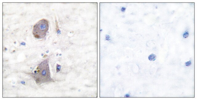
免疫细胞化学分析:代表性批次在TPA(目录号500582&524400)处理后,在人脐静脉内皮细胞(HUVEC)的膜褶中检测到PKC活化诱导的GLUT-1 Ser226磷酸化(Lee, E.E., et al. (2015).Mol.Cell.58(5):845-853)。
注意:在凝胶上样之前,通过在50°C下加热10分钟来处理裂解物样品。避免在高于60°C的温度下加热样品,会导致靶蛋白的聚集。
品質
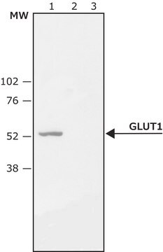
蛋白质印迹分析:2.0 µg/mL该抗体在PMA处理的HeLa细胞裂解物中检测到了Ser226磷酸化的GLUT-1。
標靶描述
其他說明
未找到合适的产品?
试试我们的产品选型工具.
儲存類別代碼
12 - Non Combustible Liquids
水污染物質分類(WGK)
WGK 1
閃點(°F)
Not applicable
閃點(°C)
Not applicable
我们的科学家团队拥有各种研究领域经验,包括生命科学、材料科学、化学合成、色谱、分析及许多其他领域.
联系技术服务部门