Skip to Content
MilliporeSigma
All Photos(2)

Key Documents

SAB4501820

Sigma-Aldrich

Anti-MAP4 antibody produced in rabbit

affinity isolated antibody

Synonym(s):

MAP 4, MTAP4, Microtubule-associated protein 4

Sign Into View Organizational & Contract Pricing


About This Item

MDL number:
UNSPSC Code:
12352203
NACRES:
NA.41

biological source

rabbit

Quality Level

conjugate

unconjugated

antibody form

affinity isolated antibody

antibody product type

primary antibodies

clone

polyclonal

form

buffered aqueous solution

mol wt

antigen 121 kDa

species reactivity

human, mouse, rat

concentration

~1 mg/mL

technique(s)

ELISA: 1:1000
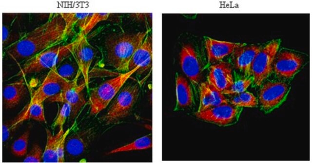
immunofluorescence: 1:100-1:500
immunohistochemistry: 1:50-1:100

NCBI accession no.

UniProt accession no.

shipped in

wet ice

storage temp.

−20°C

target post-translational modification

unmodified

Gene Information

human ... MAP4(4134)

General description

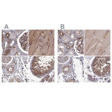
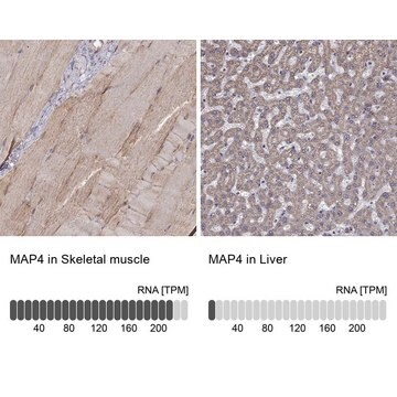
Anti-MAP4 antibody detects endogenous levels of total MAP4 protein.MAP4 (microtubule-associated protein 4) is mapped to human chromosome 3p21.31. The encoded protein is ubiquitously expressed in all cells except for the neurons. MAP4 structure contains a projection domain and a microtubule-binding (MTB) domain at the N- and C-terminal respectively.

Immunogen

The antiserum was produced against synthesized peptide derived from human MAP4.

Immunogen Range: 662-711

Biochem/physiol Actions

Microtubule-associated protein 4 (MAP4) affects microtubule assembly, stabilization and dynamics through septin interaction. MAP4 promotes apoptosis by p53 regulation. MAP4 mediates the involvement of microtubules in cell division and migration, vesicular transport or generation of primary cilia. It specifically influences mitotic spindle and centrosome position. It is known to stimulate type I IFN (interferon) signaling, which is necessary for innate immune response. MAP4 is associated with the process of invasion and metastasis of esophageal squamous cancer. It might serve as a biomarker for the same.

Features and Benefits

Evaluate our antibodies with complete peace of mind. If the antibody does not perform in your application, we will issue a full credit or replacement antibody. Learn more.

Physical form

Rabbit IgG in phosphate buffered saline (without Mg2+ and Ca2+), pH 7.4, 150mM NaCl, 0.02% sodium azide and 50% glycerol.

Disclaimer

Unless otherwise stated in our catalog or other company documentation accompanying the product(s), our products are intended for research use only and are not to be used for any other purpose, which includes but is not limited to, unauthorized commercial uses, in vitro diagnostic uses, ex vivo or in vivo therapeutic uses or any type of consumption or application to humans or animals.

Not finding the right product?  

Try our Product Selector Tool.

Storage Class

10 - Combustible liquids

wgk_germany

nwg

flash_point_f

Not applicable

flash_point_c

Not applicable


Choose from one of the most recent versions:

Certificates of Analysis (COA)

Lot/Batch Number

Don't see the Right Version?

If you require a particular version, you can look up a specific certificate by the Lot or Batch number.

Already Own This Product?

Find documentation for the products that you have recently purchased in the Document Library.

Visit the Document Library

Microtubule-Associated Protein 4 Is a Prognostic Factor and Promotes Tumor Progression in Lung Adenocarcinoma
Xia X, et al.
Disease Markers, 2018 (2018)
Chromosomal aberrations and gene expression profiles in non-small cell lung cancer
Dehan E, et al.
Lung Cancer, 56(2), 175-184 (2007)
Microtubule-associated protein 4 is an important regulator of cell invasion/migration and a potential therapeutic target in esophageal squamous cell carcinoma
Jiang YY, et al.
Oncogene, 35(37), 4846-4846 (2016)
MAP4-regulated dynein-dependent trafficking of BTN3A1 controls the TBK1-IRF3 signaling axis
Seo M, et al.
Proceedings of the National Academy of Sciences of the USA, 113(50), 14390-14395 (2016)
Microtubule associated protein-4 (MAP4) controls nanovesicle dynamics and T cell activation
Bustos-Moran E, et al.
Journal of Cell Science (2017)

Our team of scientists has experience in all areas of research including Life Science, Material Science, Chemical Synthesis, Chromatography, Analytical and many others.

Contact Technical Service