Skip to Content
MilliporeSigma
All Photos(3)

Key Documents

P5747

Sigma-Aldrich

Monoclonal Anti-Phosphoserine antibody produced in mouse

clone PSR-45, purified immunoglobulin, buffered aqueous solution

Synonym(s):

Monoclonal Anti-Phosphoserine, Phospho Ser, Phospho serine, Phospho−Ser, Phospho−serine

Sign Into View Organizational & Contract Pricing


About This Item

UNSPSC Code:
12352203
NACRES:
NA.41

biological source

mouse

Quality Level

conjugate

unconjugated

antibody form

purified immunoglobulin

antibody product type

primary antibodies

clone

PSR-45, monoclonal

form

buffered aqueous solution

packaging

antibody small pack of 25 μL

concentration

~2 mg/mL

technique(s)

dot blot: suitable
indirect ELISA: 0.3-0.6 μg/mL using phosphoserine conjugated to BSA
microarray: suitable
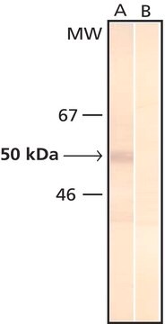
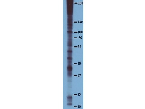
western blot: 2.5-5.0 μg/mL using total rat brain extract.

isotype

IgG1

shipped in

dry ice

storage temp.

−20°C

target post-translational modification

unmodified

Looking for similar products? Visit Product Comparison Guide

Specificity

By ELISA and dot blot, the antibody reacts specifically with phosphorylated serine, both as free amino acid or conjugated to carriers such as BSA or KLH. No cross-reactivity is observed with non-phosphorylated serine, phosphothreonine, phosphotyrosine, AMP or ATP. This antibody has been used in immunoblotting for the localization of some phosphoserine-containing proteins. Certain proteins known to contain phosphorylated serine may not be recognized by this antibody due to steric hindrance of the recognition site.

Immunogen

phosphoserine conjugated to Keyhole Limpet Hemocyanin (KLH).

Application

Applications in which this antibody has been used successfully, and the associated peer-reviewed papers, are given below.
Western Blotting (1 paper)
The antibody has been used for the detection of some phosphoserine containing proteins using immunoblotting and dot blotting.

Biochem/physiol Actions

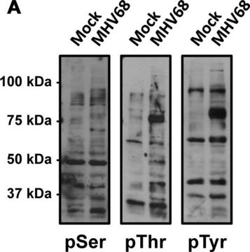
Protein phosphorylation and dephosphorylation are basic signaling mechanisms that modify protein function in eukaryotic cells. Phosphorylation is a rare posttranslational event in normal tissues, however, the abundance of phosphorylated cellular proteins increases several folds following various activation processes. The main amino acids that are phosphorylated are tyrosine, serine, or threonine (pTyr/pSer/pThr), each having specific kinases that phosphorylate them and specific phosphatases that dephosphorylate them. Mono and polyclonal antibodies directed against phosphorylated residues were generated and found useful as analytical and preparative tools by enabling the identification, quantification, and immunoaffinity isolation of phosphorylated cellular proteins.

Physical form

Solution in 0.01 M phosphate buffered saline, pH 7.4, containing 15 mM sodium azide.

Disclaimer

Unless otherwise stated in our catalog or other company documentation accompanying the product(s), our products are intended for research use only and are not to be used for any other purpose, which includes but is not limited to, unauthorized commercial uses, in vitro diagnostic uses, ex vivo or in vivo therapeutic uses or any type of consumption or application to humans or animals.

Not finding the right product?  

Try our Product Selector Tool.

Related product

Storage Class

10 - Combustible liquids

wgk_germany

nwg

flash_point_f

Not applicable

flash_point_c

Not applicable


Certificates of Analysis (COA)

Search for Certificates of Analysis (COA) by entering the products Lot/Batch Number. Lot and Batch Numbers can be found on a product’s label following the words ‘Lot’ or ‘Batch’.

Already Own This Product?

Find documentation for the products that you have recently purchased in the Document Library.

Visit the Document Library

Matteo Fornai et al.
The Journal of pharmacology and experimental therapeutics, 356(2), 434-444 (2015-11-20)
Parkinson's disease is frequently associated with gastrointestinal symptoms, mostly represented by constipation and defecatory dysfunctions. This study examined the impact of central dopaminergic denervation, induced by injection of 6-hydroxydopamine (6-OHDA) into the medial forebrain bundle, on distal colonic excitatory cholinergic
An integrated assessment of histopathological changes of the enteric neuromuscular compartment in experimental colitis
Ippolito C, et al.
Journal of Cellular and Molecular Medicine, 19(2), 485-500 (2015)
M Zhao et al.
FEBS letters, 509(3), 405-412 (2001-12-26)
B-cell leukemia/lymphoma 2 (Bcl-2) blocks oxidant-induced apoptosis at least partly by stabilizing lysosomes. Here we report that phosphorylation of Bcl-2 may be required for these protective effects. J774 cells overexpressing wild-type Bcl-2 resist oxidant-induced lysosomal leak as well as apoptosis
P R Murphy et al.
Endocrinology, 142(1), 81-88 (2001-01-06)
We recently reported that the rat Nb2 T lymphoma cells expressed messenger RNAs (mRNAs) encoding both fibroblast growth factor-2 (FGF-2) and the FGF receptor, suggesting possible paracrine and/or autocrine roles for FGF-2 in lymphoma cell function. We have also shown
Mechanisms of specificity in protein phosphorylation
Ubersax JA and Ferrell Jr JE
Nature Reviews in Molecular and Cell Biology, 8(7), 530-530 (2007)

Our team of scientists has experience in all areas of research including Life Science, Material Science, Chemical Synthesis, Chromatography, Analytical and many others.

Contact Technical Service