Skip to Content
MilliporeSigma
All Photos(2)

Key Documents

L8793

Sigma-Aldrich

Anti-MAP1LC3A antibody

rabbit polyclonal

Synonym(s):

Anti-MAP1 light chain 3-like protein 1, Anti-MAP1A/1B light chain 3 A, Anti-MAP1A/MAP1B LC3 A, Anti-MAP1ALC3, Anti-MAP1BLC3, Anti-MAP1LC3A, Anti-Microtubule-associated protein 1 light chain 3 alpha

Sign Into View Organizational & Contract Pricing


About This Item

UNSPSC Code:
12352203
NACRES:
NA.41

Product Name

Anti-LC3A antibody produced in rabbit, ~1 mg/mL, affinity isolated antibody, buffered aqueous solution

biological source

rabbit

Quality Level

conjugate

unconjugated

antibody form

affinity isolated antibody

antibody product type

primary antibodies

clone

polyclonal

form

buffered aqueous solution

mol wt

antigen 16-18 kDa

species reactivity

mouse, human, rat

concentration

~1 mg/mL

technique(s)

immunoprecipitation (IP): 5-10 μg using extracts of human U87 cells
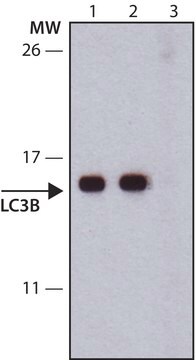
western blot: 0.5-1 μg/mL using whole extracts of rat and mouse brain

UniProt accession no.

shipped in

dry ice

storage temp.

−20°C

target post-translational modification

unmodified

Gene Information

General description

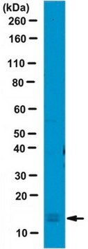
Microtubule-associated proteins 1A/1B light chain 3 or MAP1LC3 is a cellular protein that is involved in the formation of autophagosomes . MAP1LC3 genes have three known human isoforms, namely, MAP1LC3A, MAP1LC3B and MAP1LC3C. These isoforms have different responses to amino acid starvation and varied sub-cellular localization patterns . Anti-LC3A antibody reacts with human and rat LC3A-I (approx. 18 kDa) and LC3A-II (approx. 16 kDa) by immunoblotting and immunoprecipitation. The antibody is also specific for mouse LC3A-I (approx. 18 kDa). Specific cell treatment and/or long exposure may be required for the detection of mouse LC3A-II. The immunizing peptide specifically inhibits the detection of the LC3A bands by immunoblotting.

Specificity

Anti-LC3A antibody recognizes human and rat microtubule-associated protein light chain 3-A (LC3A-I) and LC3A-II by immunoblotting and immunoprecipitation (~18 kDa and ~16 kDa, respectively) and mouse LC3A-I (~18 kDa).

Immunogen

Synthetic peptide corresponding to amino acids of human LC3A isoform α, conjugated to KLH via C-terminal cysteine residue. The corresponding sequence is identical in rat and mouse.

Application

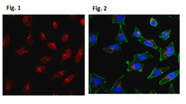
Anti-LC3A antibody produced in rabbit has been used in
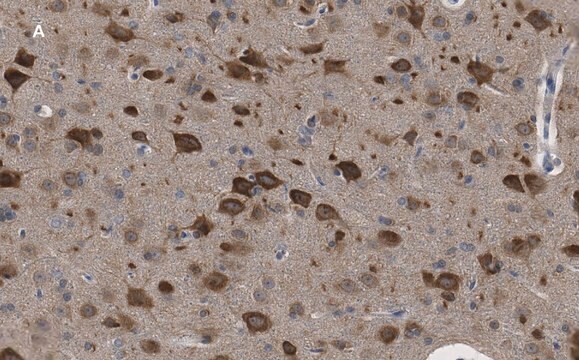
  • immunohistochemistry
  • immunostaining
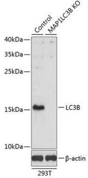
  • western blotting

Biochem/physiol Actions

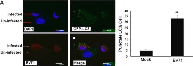
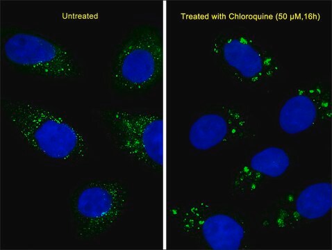
Microtubule-associated protein light chain 3-I (LC3-I) is modified to a membrane-bound form, LC3-II (a LC3-phospholipid conjugate), by mammalian autophagy related 7 (Atg7) and Atg3, which are E1- and E2-like enzymes, respectively. The amount of LC3 II correlates with the extent of autophagosome formation. LC3A can also be as an autophagosomal marker.

Physical form

Solution in 0.01 M phosphate buffered saline, pH 7.4, containing 15 mM sodium azide.

Storage and Stability

For continuous use, store at 2-8 °C for up to one month. For extended storage, freeze in working aliquots. Repeated freezing and thawing, or storage in “frost-free” freezers, is not recommended. If slight turbidity occurs upon prolonged storage, clarify the solution by centrifugation before use. Working dilutions should be discarded if not used within 12 hours.

Disclaimer

Unless otherwise stated in our catalog or other company documentation accompanying the product(s), our products are intended for research use only and are not to be used for any other purpose, which includes but is not limited to, unauthorized commercial uses, in vitro diagnostic uses, ex vivo or in vivo therapeutic uses or any type of consumption or application to humans or animals.

Not finding the right product?  

Try our Product Selector Tool.

Storage Class

10 - Combustible liquids

wgk_germany

WGK 3

flash_point_f

Not applicable

flash_point_c

Not applicable

ppe

Eyeshields, Gloves, multi-purpose combination respirator cartridge (US)


Choose from one of the most recent versions:

Certificates of Analysis (COA)

Lot/Batch Number

Don't see the Right Version?

If you require a particular version, you can look up a specific certificate by the Lot or Batch number.

Already Own This Product?

Find documentation for the products that you have recently purchased in the Document Library.

Visit the Document Library

The role of lysosomes and autophagosomes in frontotemporal lobar degeneration
Bain HDC, et al.
Neuropathology and Applied Neurobiology, 45(3), 244-261 (2019)
Xuekai Zhang et al.
Alzheimer's research & therapy, 6(9), 78-78 (2015-01-27)
Many neurodegenerative diseases are characterised by accumulations of misfolded proteins that can colocalise with chaperone proteins (for example, heat shock protein 27 (HSP27)), which might act as modulators of protein aggregation. The role of HSP27 in the pathogenesis of neurodegenerative
Expression of one important chaperone protein, heat shock protein 27, in neurodegenerative diseases
Zhang X, et al.
Alzheimer's Research & Therapy, 6(9), 78-78 (2014)
Rossana C N Melo et al.
Nature protocols, 9(10), 2382-2394 (2014-09-12)
Precise immunolocalization of proteins within a cell is central to understanding cell processes and functions such as intracellular trafficking and secretion of molecules during immune responses. Here we describe a protocol for ultrastructural detection of proteins in leukocytes. The method
João Cláudio Damasceno-Sá et al.
Memorias do Instituto Oswaldo Cruz, 116, e200417-e200417 (2021-03-18)
Toxoplasma gondii causes toxoplasmosis and is controlled by activated macrophages. However, infection of macrophages by tachyzoites induces TGF-β signaling (TGF-s) inhibiting nitric oxide (NO) production. NO inhibition may be a general escape mechanism of distinct T. gondii strains. To evaluate

Our team of scientists has experience in all areas of research including Life Science, Material Science, Chemical Synthesis, Chromatography, Analytical and many others.

Contact Technical Service