Skip to Content
MilliporeSigma
All Photos(3)

Key Documents

AV42202

Sigma-Aldrich

Anti-ACTN4 (AB2) antibody produced in rabbit

IgG fraction of antiserum

Synonym(s):

Anti-Actinin, α4, Anti-DKFZp686K23158, Anti-FSGS, Anti-FSGS1

Sign Into View Organizational & Contract Pricing


About This Item

UNSPSC Code:
12352203
NACRES:
NA.41

biological source

rabbit

Quality Level

conjugate

unconjugated

antibody form

IgG fraction of antiserum

antibody product type

primary antibodies

clone

polyclonal

form

buffered aqueous solution

mol wt

100 kDa

species reactivity

bovine, dog, guinea pig, horse, rat, human, mouse, rabbit

concentration

0.5 mg - 1 mg/mL

technique(s)

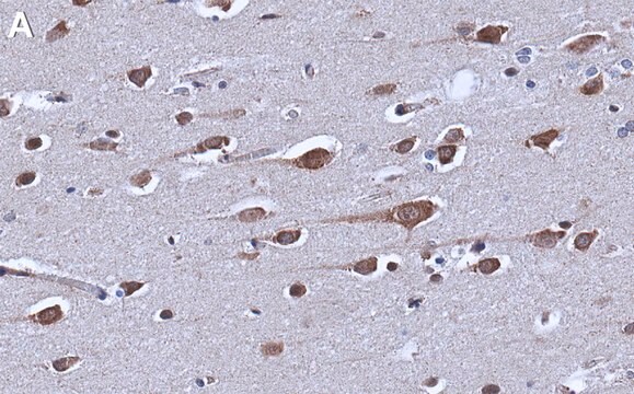
immunohistochemistry: suitable
western blot: suitable

NCBI accession no.

UniProt accession no.

shipped in

wet ice

storage temp.

−20°C

target post-translational modification

unmodified

Gene Information

human ... ACTN4(81)

Immunogen

Synthetic peptide directed towards the N terminal region of human ACTN4

Biochem/physiol Actions

Alpha actinins belong to the spectrin superfamily which represents a diverse group of cytoskeletal proteins, including the alpha and beta spectrins and dystrophins. Alpha actinin is an actin-binding protein with multiple roles in different cell types. In nonmuscle cells, the cytoskeletal isoform is found along microfilament bundles and adherens-type junctions, where it is involved in binding actin to the membrane. In contrast, skeletal, cardiac, and smooth muscle isoforms are localized to the Z-disc and analogous dense bodies, where they help anchor the myofibrillar actin filaments. ACTN4 is a nonmuscle, alpha actinin isoform which is concentrated in the cytoplasm, and thought to be involved in metastatic processes. Mutations in its gene have been associated with focal and segmental glomerulosclerosis.Alpha actinins belong to the spectrin gene superfamily which represents a diverse group of cytoskeletal proteins, including the alpha and beta spectrins and dystrophins. Alpha actinin is an actin-binding protein with multiple roles in different cell types. In nonmuscle cells, the cytoskeletal isoform is found along microfilament bundles and adherens-type junctions, where it is involved in binding actin to the membrane. In contrast, skeletal, cardiac, and smooth muscle isoforms are localized to the Z-disc and analogous dense bodies, where they help anchor the myofibrillar actin filaments. This gene encodes a nonmuscle, alpha actinin isoform which is concentrated in the cytoplasm, and thought to be involved in metastatic processes. Mutations in this gene have been associated with focal and segmental glomerulosclerosis.

Sequence

Synthetic peptide located within the following region: LIHRHRPELIEYDKLRKDDPVTNLNNAFEVAEKYLDIPKMLDAEDIVNTA

Physical form

Purified antibody supplied in 1x PBS buffer with 0.09% (w/v) sodium azide and 2% sucrose.

Disclaimer

Unless otherwise stated in our catalog or other company documentation accompanying the product(s), our products are intended for research use only and are not to be used for any other purpose, which includes but is not limited to, unauthorized commercial uses, in vitro diagnostic uses, ex vivo or in vivo therapeutic uses or any type of consumption or application to humans or animals.

Not finding the right product?  

Try our Product Selector Tool.

Storage Class

10 - Combustible liquids

wgk_germany

WGK 3

flash_point_f

Not applicable

flash_point_c

Not applicable


Certificates of Analysis (COA)

Search for Certificates of Analysis (COA) by entering the products Lot/Batch Number. Lot and Batch Numbers can be found on a product’s label following the words ‘Lot’ or ‘Batch’.

Already Own This Product?

Find documentation for the products that you have recently purchased in the Document Library.

Visit the Document Library

Carole L Moncman et al.
Experimental cell research, 319(3), 23-31 (2012-11-24)
Extraocular muscles are a unique subset of striated muscles. During postnatal development, the extraocular muscles undergo a number of myosin isoform transitions that occur between postnatal day P10 (P10) and P15. These include: (1) loss of embryonic myosin from the

Our team of scientists has experience in all areas of research including Life Science, Material Science, Chemical Synthesis, Chromatography, Analytical and many others.

Contact Technical Service