Skip to Content
MilliporeSigma
All Photos(1)

Key Documents

380R-2

Sigma-Aldrich

Arginase-1 (EP261) Rabbit Monoclonal Primary Antibody

Sign Into View Organizational & Contract Pricing


About This Item

UNSPSC Code:
12352203

biological source

rabbit

Quality Level

100
500

conjugate

unconjugated

antibody form

culture supernatant

antibody product type

primary antibodies

clone

EP261, monoclonal

description

For In Vitro Diagnostic Use in Select Regions

form

buffered aqueous solution

species reactivity

human

packaging

vial of 0.1 mL concentrate (380R-24)
vial of 0.1 mL concentrate Research Use Only (380R-24-RUO)
vial of 0.5 mL concentrate (380R-25)
vial of 1.0 mL concentrate (380R-26)
vial of 1.0 mL concentrate Research Use Only (380R-26-RUO)
vial of 1.0 mL pre-dilute Research Use Only (380R-27-RUO)
vial of 1.0 mL pre-dilute ready-to-use (380R-27)
vial of 7.0 mL pre-dilute ready-to-use (380R-28)
vial of 7.0 mL pre-dilute ready-to-use Research Use Only (380R-28-RUO)

manufacturer/tradename

Cell Marque

technique(s)

immunohistochemistry (formalin-fixed, paraffin-embedded sections): 1:50-1:200 (concentrated)

isotype

IgG

control

hepatocellular carcinoma, normal liver

shipped in

wet ice

storage temp.

2-8°C

visualization

cytoplasmic, nuclear

Gene Information

human ... ARG1(383)

General description

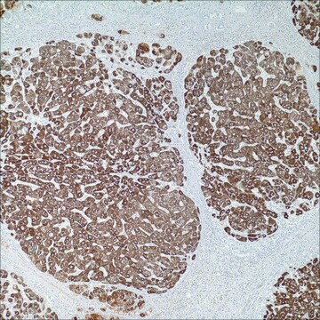
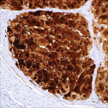
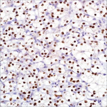
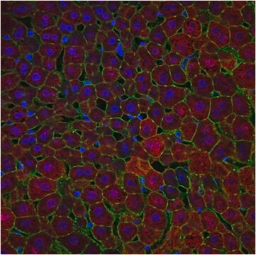
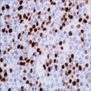
Arginase-1 is a key urea cycle metalloenzyme that has demonstrated expression in normal human liver with a high degree of specificity. Hepatocellular carcinoma (HCC) is the most common primary malignant tumor of the liver accounting for an estimated 70%-85% of total liver cancers worldwide. Diagnostic pitfalls exist in the morphologic distinction of HCC from other hepatocellular and non-hepatocellular lesions. In difficult or equivocal cases, the application of immunohistochemical (IHC) panels has been shown to aid in the distinction of benign and malignant liver lesions. In sections of normal liver, anti-arginase-1 produced strong, diffuse cytoplasmic reactivity in all hepatocytes throughout the lobule. In some cases, patchy nuclear reactivity is also evident in hepatocytes along with the cytoplasmic reactivity. Reactivity is not observed in bile duct epithelial cells, sinusoidal endothelial cells, Kupffer cells, or vascular endothelial cells. In sections of HCC, anti-arginase-1 produces cytoplasmic or cytoplasmic plus nuclear reactivity.

Quality


IVD

IVD

IVD

RUO

Linkage

Arginase-1 Positive Control Slides, Product No. 380S, are available for immunohistochemistry (formalin-fixed, paraffin-embedded sections).

Physical form

Solution in Tris Buffer, pH 7.3-7.7, with 1% BSA and <0.1% Sodium Azide.

Preparation Note

Download the IFU specific to your product lot and format
Note: This requires a keycode which can be found on your packaging or product label.

Download the latest released IFU
Note: This IFU may not apply to your specific product lot.

Other Notes

For Technical Service please contact: 800-665-7284 or email: service@cellmarque.com

Legal Information

Cell Marque is a trademark of Merck KGaA, Darmstadt, Germany

Not finding the right product?  

Try our Product Selector Tool.

Storage Class

12 - Non Combustible Liquids

wgk_germany

WGK 2

flash_point_f

Not applicable

flash_point_c

Not applicable


Certificates of Analysis (COA)

Search for Certificates of Analysis (COA) by entering the products Lot/Batch Number. Lot and Batch Numbers can be found on a product’s label following the words ‘Lot’ or ‘Batch’.

Already Own This Product?

Find documentation for the products that you have recently purchased in the Document Library.

Visit the Document Library

R L Zimmerman et al.
Cancer, 93(4), 288-291 (2001-08-17)
Diagnosing liver tumors by fine-needle aspiration biopsy is safe and accurate. However, there are cases that prove diagnostically difficult. Traditionally, immunostains for alpha-fetoprotein and polyclonal carcinoembryonic antigen have been used to distinguish adenocarcinomas from hepatocellular carcinomas (HCCs). In poorly differentiated
Nehal A Radwan et al.
Diagnostic pathology, 7, 149-149 (2012-11-01)
The ability to distinguish hepatocellular carcinoma (HCC) from metastatic carcinoma (MC) involving the liver and cholangiocarcinoma (CC) by immunohistochemistry has been limited by the lack of a reliable positive marker for hepatocellular differentiation. Arginase-1 is a marker for HCC recently
T H Niemann et al.
Cancer, 87(5), 295-298 (1999-10-28)
Fine-needle aspiration biopsy (FNAB) is frequently used to diagnose mass lesions in the liver. Differentiating metastatic adenocarcinoma from primary hepatocellular carcinoma can be difficult. Despite a number of morphologic criteria, there remain occasional cases in which the cytologic features fail
Alexandre Sherlley Casimiro Onofre et al.
Cancer, 111(4), 259-268 (2007-06-15)
Difficulties with cytologic diagnoses on fine-needle aspiration cytology (FNAC) of the liver can be overcome by the application of immunocytochemical panels applied on smears. The aim of the current study was to analyze the performance of a panel of monoclonal
Ahmedin Jemal et al.
CA: a cancer journal for clinicians, 61(2), 69-90 (2011-02-08)
The global burden of cancer continues to increase largely because of the aging and growth of the world population alongside an increasing adoption of cancer-causing behaviors, particularly smoking, in economically developing countries. Based on the GLOBOCAN 2008 estimates, about 12.7

Our team of scientists has experience in all areas of research including Life Science, Material Science, Chemical Synthesis, Chromatography, Analytical and many others.

Contact Technical Service