Skip to Content
MilliporeSigma
All Photos(1)

Key Documents

130M-9

Sigma-Aldrich

CD30 (Ber-H2) Mouse Monoclonal Antibody

Sign Into View Organizational & Contract Pricing


About This Item

UNSPSC Code:
12352200
NACRES:
NA.41

biological source

mouse

Quality Level

100
500

conjugate

unconjugated

antibody form

culture supernatant

antibody product type

primary antibodies

clone

Ber-H2, monoclonal

description

For In Vitro Diagnostic Use in Select Regions (See Chart)

form

buffered aqueous solution

species reactivity

human

packaging

vial of 0.1 mL concentrate (130M-94)
vial of 0.5 mL concentrate (130M-95)
bottle of 1.0 mL predilute (130M-97)
vial of 1.0 mL concentrate (130M-96)
bottle of 7.0 mL predilute (130M-98)

manufacturer/tradename

Cell Marque

technique(s)

immunohistochemistry (formalin-fixed, paraffin-embedded sections): 1:50-1:200

isotype

IgG1κ

control

classic Hodgkin lymphoma

shipped in

wet ice

storage temp.

2-8°C

visualization

membranous

Gene Information

human ... TNFRSF8(943)

General description

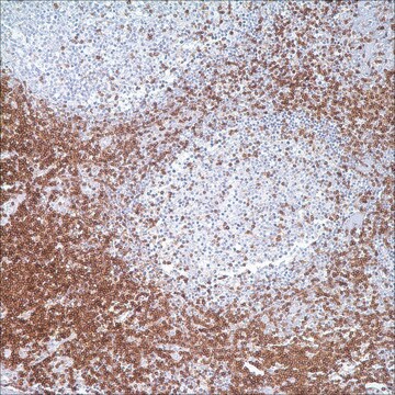
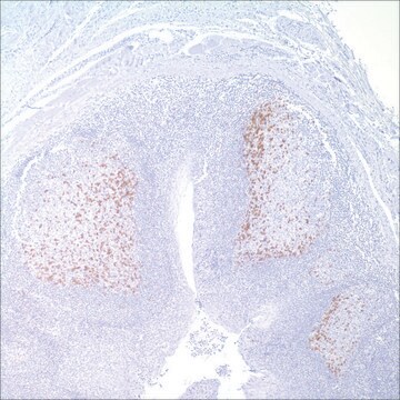
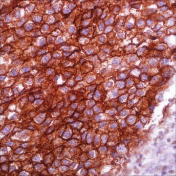
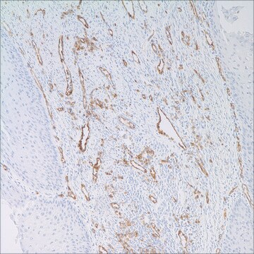
Anti-CD30 detects a formalin-resistant epitope that is expressed by Reed-Sternberg cells in classic Hodgkin lymphoma, the majority of anaplastic large cell lymphomas, primary cutaneous CD30 positive T-cell lymphoproliferative disorders, and in embryonal carcinomas. Occasionally diffuse large B-cell lymphoma stains with this antibody. This antibody also stains plasma cells in paraffin-embedded tissue. However, reactive immunoblasts are immunoreactive with this antibody. The staining pattern of anti-CD30 in lymphoma or embryonal carcinoma is different, with the former being membranous and exhibiting Golgi zone accentuation in location, and the latter being membranous only.

Quality


IVD

IVD

IVD

RUO

Linkage

CD30 Positive Control Slides, Product No. 130S, are available for immunohistochemistry (formalin-fixed, paraffin-embedded sections).

Physical form

Solution in Tris Buffer, pH 7.3-7.7, with 1% BSA and <0.1% Sodium Azide

Preparation Note

Download the IFU specific to your product lot and formatNote: This requires a keycode which can be found on your packaging or product label.

Other Notes

For Technical Service please contact: 800-665-7284 or email: service@cellmarque.com

Legal Information

Cell Marque is a trademark of Merck KGaA, Darmstadt, Germany

Not finding the right product?  

Try our Product Selector Tool.


Certificates of Analysis (COA)

Search for Certificates of Analysis (COA) by entering the products Lot/Batch Number. Lot and Batch Numbers can be found on a product’s label following the words ‘Lot’ or ‘Batch’.

Already Own This Product?

Find documentation for the products that you have recently purchased in the Document Library.

Visit the Document Library

Rabban JT, et al.
Diagnostic Immunohistochemistry: Theranostic and Genomic Applications, 702-702 (2014)
David H George et al.
The American journal of surgical pathology, 27(4), 487-493 (2003-03-27)
Anaplastic large cell lymphoma (ALCL) rarely occurs in the central nervous system. Although defined by its composition of large, pleomorphic, CD30-positive lymphocytes, ALCL is heterogeneous. Most are T cell but some are null cell. Most but not all have a
R Schwarting et al.
Blood, 74(5), 1678-1689 (1989-10-01)
The production and characterization of a monoclonal antibody (MoAb) designated Ber-H2, directed against a new epitope of the Ki-1 (CD30) antigen, are described. In comparison with the formerly reported Ki-1 MoAb whose reactivity with Hodgkin and Reed-Sternberg (H-RS) cells in
Cyrus V Hedvat et al.
Human pathology, 33(10), 968-974 (2002-10-24)
The immunohistochemical analysis of lymphoid neoplasms has led to refined classification schemes based on the profile of antigen expression and correlation with morphological, cytogenetic, molecular, and clinical features. Tissue microarrays (TMAs) are a powerful tool to rapidly characterize the phenotypic

Our team of scientists has experience in all areas of research including Life Science, Material Science, Chemical Synthesis, Chromatography, Analytical and many others.

Contact Technical Service