Skip to Content
MilliporeSigma
All Photos(2)

Key Documents

MABN845

Sigma-Aldrich

Anti-Neurophysin 2/NP-AVP Antibody, clone PS 41

clone PS 41, from mouse

Synonym(s):

Vasopressin-neurophysin 2-copeptin, AVP-NPII, Arg-vasopressin, Arginine-vasopressin, Neurophysin 2, Neurophysin-I, Copeptin, Neurophysin 2/NP-AVP

Sign Into View Organizational & Contract Pricing


About This Item

UNSPSC Code:
12352203
eCl@ss:
32160702
NACRES:
NA.41

biological source

mouse

Quality Level

antibody form

purified immunoglobulin

antibody product type

primary antibodies

clone

PS 41, monoclonal

species reactivity

rat

technique(s)

ELISA: suitable
electron microscopy: suitable
immunohistochemistry: suitable
immunoprecipitation (IP): suitable
radioimmunoassay: suitable
western blot: suitable

isotype

IgG2bκ

NCBI accession no.

UniProt accession no.

shipped in

wet ice

target post-translational modification

unmodified

Gene Information

rat ... Avp(24221)

General description

The neurophysin 1 and 2 (NPI and NPII) are carriers of peptide hormone oxytocin (OT) and vasopressin (AVP), respectively. Each NP and its associated hormone peptide are synthesized as one precursor protein, OT-NPI (UniProt P01179) encoded by the Oxt (or Ot) gene or AVP-NPII (UniProt P01186) encoded by the Avp gene in rat species. Post-translational cleavages remove the signal peptide and process the precursor into mature OT and NPI (aa 20-28 and aa 32-135, respectively, of OT-NPI) or AVP and NPII (aa 24-32 and aa 36-128, respectively, of AVP-NPII). OT is a potent hormone and neurotransmitter secreted within the pituitary gland. OT exerts its biological activity through its receptor OXTR (also known as OTR). OT is used clinically for labor induction and may exhibit therapeutic potential for treating social anxiety disorders. AVP functions primarily as an anti-diuretic and regulates the concentration of water, glucose, and salts in blood. AVP is secreted from the posterior pituitary, but exerts its biological activity in the kidney via the G protein-coupled receptor AVPR2 (V2R).

Specificity

Clone PS 41 reacts with Vasopressin-neurophysin 2-copeptin precursor, neurophysin 2, but not Arg-vasopressin, copeptin, oxytocin, or neurophysin 1.

Immunogen

Epitope: Neurophysin 2
KLH-conjugated acid soluble extract of rat (Sprague-Dawley) intermediate lobes.

Application

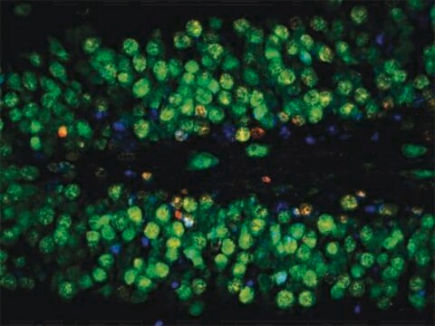
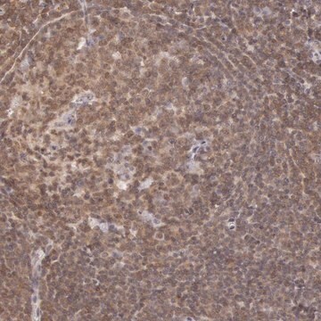
Immunohistochemistry Analysis: A 1:1,000 dilution from a representative lot detected Neurophysin 2/NP-AVP in rat pituitary tissue.
Immunohistochemistry Analysis: A representative lot detected Neurophysin 2/NP-AVP immunoreactivity in the hypothalamus of developing rats as early as embryonic day (E16) using coronal vibratome sections. (Witnall, M.H., et al.(1985). J Neurosci. 5(1):98-109).
Radioimmunoassay Analysis: Target reactivity of a representative lot was confirmed by RIA using plates coated with purified rat Neurophysin/NP and 125I-labelled secondary antibody (Ben-Barak, Y., et al. (1985). J Neurosci. 5(1):81-97).
Electron Microscopy Analysis: A representative lot immunostained granulated neurosecretory vesicles (NSVs) in all axons, Herring bodies, and terminals by EM using rat posterior pituitary LR White sections (Ben-Barak, Y., et al. (1985). J Neurosci. 5(1):81-97).
Immunoprecipitation Analysis: A representative lot immunoprecipitated radiolabelled neurophysin 2, but not neurophysin 1 (Ben-Barak, Y., et al. (1985). J Neurosci. 5(1):81-97).
ELISA Analysis: A representative lot detected neurophysin 2 immunoreactivity in rat neurophysin preparations by non-sandwich ELISA using either 125I-labelled or HRP-conjugated secondary antibody (Ben-Barak, Y., et al. (1985). J Neurosci. 5(1):81-97).
Research Category
Neuroscience
Research Sub Category
Developmental Neuroscience
This Anti-Neurophysin 2/NP-AVP Antibody, clone PS 41 is validated for use in Western Blotting and Immunohistochemistry, Radioimmunoassay, Electron Microscopy, Immunoprecipitation and ELISA for the detection of Neurophysin 2/NP-AVP.

Quality

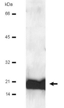
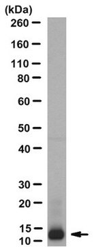
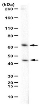
Evaluated by Western Blotting in rat pituitary tissue lysate.

Western Blotting Analysis: 0.5 µg/mL of this antibody detected Neurophysin 2/NP-AVP in 10 µg of rat pituitary tissue lysate.

Target description

~15 kDa observed

Physical form

Format: Purified
Protein G Purified
Purified mouse monoclonal IgG2bκ antibody in buffer containing 0.1 M Tris-Glycine (pH 7.4), 150 mM NaCl with 0.05% sodium azide.

Storage and Stability

Stable for 1 year at 2-8°C from date of receipt.

Other Notes

Concentration: Please refer to lot specific datasheet.

Disclaimer

Unless otherwise stated in our catalog or other company documentation accompanying the product(s), our products are intended for research use only and are not to be used for any other purpose, which includes but is not limited to, unauthorized commercial uses, in vitro diagnostic uses, ex vivo or in vivo therapeutic uses or any type of consumption or application to humans or animals.

Not finding the right product?  

Try our Product Selector Tool.

Storage Class

12 - Non Combustible Liquids

wgk_germany

WGK 1

flash_point_f

Not applicable

flash_point_c

Not applicable


Certificates of Analysis (COA)

Search for Certificates of Analysis (COA) by entering the products Lot/Batch Number. Lot and Batch Numbers can be found on a product’s label following the words ‘Lot’ or ‘Batch’.

Already Own This Product?

Find documentation for the products that you have recently purchased in the Document Library.

Visit the Document Library

Neurophysin in the hypothalamo-neurohypophysial system. II. Immunocytochemical studies of the ontogeny of oxytocinergic and vasopressinergic neurons.
Whitnall, MH; Key, S; Ben-Barak, Y; Ozato, K; Gainer, H
The Journal of Neuroscience null
Mingfeng Zhou et al.
Neuroendocrinology (2021-11-12)
Hypothalamic injury causes several complicated neuroendocrine-associated disorders, such as water-electrolyte imbalance, obesity, and hypopituitarism. Among these, central diabetes insipidus (CDI), characterized by polyuria, polydipsia, low urine specific gravity, and deficiency of arginine vasopressin contents, is a typical complication after hypothalamic
Marie Habart et al.
STAR protocols, 4(1), 101968-101968 (2023-01-05)
Here, we present an optimized iDISCO+ protocol combining tissue clearing and light sheet microscopy to map the postnatal development of oxytocin and vasopressin neurons in mouse hypothalamus. We describe tissue preparation, immunostaining, clearing, and imaging. We then detail how to
Neurophysin in the hypothalamo-neurohypophysial system. I. Production and characterization of monoclonal antibodies.
Ben-Barak, Y; Russell, JT; Whitnall, MH; Ozato, K; Gainer, H
The Journal of Neuroscience null

Our team of scientists has experience in all areas of research including Life Science, Material Science, Chemical Synthesis, Chromatography, Analytical and many others.

Contact Technical Service