Skip to Content
MilliporeSigma
All Photos(1)

Key Documents

MABN162

Sigma-Aldrich

Anti-Tau, a.a. 210-241 Antibody, clone Tau-5

clone TAU-5, from mouse

Synonym(s):

Microtubule-associated protein tau, Neurofibrillary tangle protein, Paired helical filament-tau, PHF-tau

Sign Into View Organizational & Contract Pricing


About This Item

UNSPSC Code:
12352203
eCl@ss:
32160702
NACRES:
NA.41

biological source

mouse

Quality Level

antibody form

purified immunoglobulin

antibody product type

primary antibodies

clone

TAU-5, monoclonal

species reactivity

human, rat

species reactivity (predicted by homology)

bovine (immunogen homology)

technique(s)

western blot: suitable

isotype

IgG1κ

NCBI accession no.

UniProt accession no.

shipped in

wet ice

target post-translational modification

unmodified

Gene Information

human ... MAPT(4137)
mouse ... Mapt(281296)
rat ... Mapt(29477)

General description

Tau is a neuronal protein implicated in several neurodegenerative disorders, among which Alzheimer’s disease, Picks’s disease, and frontotemporal dementia are most widely studied. Tau interacts extensively with the cytoskeleton via its microtubule-binding domain and is involved in the assembly and stabilization of microtubules. Tau also contains proline rich domains and multiple phoshorylation sites which are targeted by a wide variety of kinases including JNK, MAPK, GSK3, PKA, PKC, and CAMKII. In disease conditions, Tau is hyperphosphorylated and insoluble and occurs mainly in the form of toxic neurofibrillary tangles that destabilizes the cytoskeleton, and disrupts axonal transport and intracellular signaling networks. Neurodegeneration resulting from accumulation of defective tau proteins is marked by a range of cognitive deficits, among other debilitating symptoms.

Immunogen

Purified bovine Tau

Application

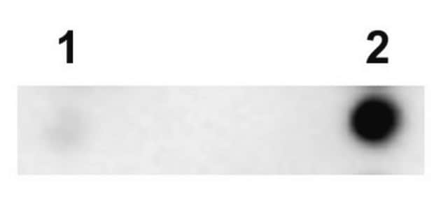
Detect Tau, a.a. 210-241, clone Tau-5 using this Anti-Tau, a.a. 210-241 Antibody, clone Tau-5 validated for use in Western Blotting.
Research Category
Neuroscience
Research Sub Category
Neurodegenerative Diseases

Quality

Purified by Western Blot in rat cortex tissue lysate.

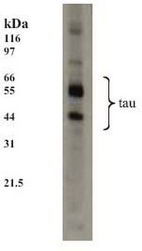
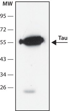
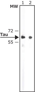
Western Blot Analysis: 0.1 µg/mL of this antibody recognized Tau in 10 µg of rat cortex tissue lysate.

Target description

~48 kDa observed. This antibody is expected to recognize all phosphorylated and nonphosphorylated isoforms of Tau at molecular weights of ~45-68 kDa

Linkage

Replaces: MABN361; 577801

Physical form

Format: Purified
Protein G Purified
Purified mouse monoclonal IgG1κ in buffer containing PBS without preservatives.

Storage and Stability

Stable for 1 year at -20°C from date of receipt.
Handling Recommendations: Upon receipt and prior to removing the cap, centrifuge the vial and gently mix the solution. Aliquot into microcentrifuge tubes and store at -20°C. Avoid repeated freeze/thaw cycles, which may damage IgG and affect product performance.

Analysis Note

Control
Rat cortex tissue lysate

Other Notes

Concentration: Please refer to the Certificate of Analysis for the lot-specific concentration.

Disclaimer

Unless otherwise stated in our catalog or other company documentation accompanying the product(s), our products are intended for research use only and are not to be used for any other purpose, which includes but is not limited to, unauthorized commercial uses, in vitro diagnostic uses, ex vivo or in vivo therapeutic uses or any type of consumption or application to humans or animals.

Not finding the right product?  

Try our Product Selector Tool.

Storage Class

12 - Non Combustible Liquids

wgk_germany

WGK 2

flash_point_f

Not applicable

flash_point_c

Not applicable


Certificates of Analysis (COA)

Search for Certificates of Analysis (COA) by entering the products Lot/Batch Number. Lot and Batch Numbers can be found on a product’s label following the words ‘Lot’ or ‘Batch’.

Already Own This Product?

Find documentation for the products that you have recently purchased in the Document Library.

Visit the Document Library

Lucia Buccarello et al.
Oncotarget, 8(47), 83038-83051 (2017-11-16)
Recently a range of ocular manifestations such as retinal and lens amyloid-beta accumulation and retinal nerve fiber layer loss have been proposed as potential biomarkers in Alzheimer disease (AD). The TgCRND8 mouse model of AD exhibits age-dependent amyloid β (Aβ)

Our team of scientists has experience in all areas of research including Life Science, Material Science, Chemical Synthesis, Chromatography, Analytical and many others.

Contact Technical Service