MABE251
Anti-5-hydroxymethylcytosine (5hmC) Antibody, clone HMC 31
clone HMC 31, from mouse
Sign Into View Organizational & Contract Pricing
All Photos(2)
About This Item
UNSPSC Code:
12352203
eCl@ss:
32160702
NACRES:
NA.41
Recommended Products
biological source
mouse
Quality Level
antibody form
purified immunoglobulin
antibody product type
primary antibodies
clone
HMC 31, monoclonal
species reactivity (predicted by homology)
all
technique(s)
ELISA: suitable
dot blot: suitable
methylated DNA immunoprecipitation (MeDIP): suitable
isotype
IgG1κ
shipped in
wet ice
target post-translational modification
unmodified
General description
5-Hydroxymethylcytosine is a DNA pyrimidine nitrogen base formed from the enzymatic conversion of 5-methylcytosine into 5-hydroxymethylcytosine by the TET family of iron-dependent oxygenases. Data suggests that every mammilian cell contains 5-hydroxymethylcytosine, but the levels vary depending on the cell type; data also suggests that levels of 5-hydroxymethylcytosine increases with age. The highest levels are found in neuronal cells of the central nervous system and certain mammalian tissues such as mouse Purkinje and granule neurons. Although the exact function has not been fully elucidated, studies suggest that 5-hydroxymethlcytosine may regulate gene expression or initiate DNA demethylation.
Specificity
This antibody recognizes the 5-hydroxymethylcytosine of 5-Hydroxymethylcytosine DNA.
Immunogen
BSA-conjugated 5-hydroxymethylcytosine.
Epitope: 5-hydroxymethylcytosine
Application
Detect 5-hydroxymethylcytosine (5hmC) with Anti-5-hydroxymethylcytosine (5hmC) Antibody, clone HMC 31 (Mouse Monoclonal Antibody), that has been shown to work in DB, MeDIP, ELISA.
ELISA Analysis: A representative lot detected 5-hydroxymethylcytosine in ELISA.
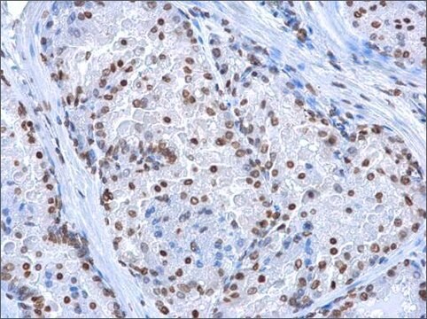
Methylated DNA Immunoprecipitation (MeDIP) Analysis: A representative lot immunoprecipitated 5-hydroxymethylcytosine in ChIP.
Methylated DNA Immunoprecipitation (MeDIP) Analysis: A representative lot immunoprecipitated 5-hydroxymethylcytosine in ChIP.
Research Category
Epigenetics & Nuclear Function
Epigenetics & Nuclear Function
Research Sub Category
Chromatin Biology
Chromatin Biology
Quality
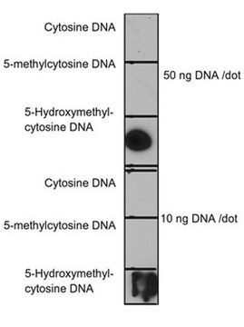
Evaluated by Dot Blot in unmodified and modified cytosine DNA.
Dot Blot Analysis: 0.1 µg/mL of this antibody detected 5-hydroxymethylcytosine in 10 ng and 50 ng of unmodified and modified cytosine DNA.
Dot Blot Analysis: 0.1 µg/mL of this antibody detected 5-hydroxymethylcytosine in 10 ng and 50 ng of unmodified and modified cytosine DNA.
Target description
This antibody detects 5-hydroxymethylcytosine.
Physical form
Format: Purified
Protein G Purified
Purified mouse monoclonal IgG1κ in buffer containing 0.1 M Tris-Glycine (pH 7.4), 150 mM NaCl with 0.05% sodium azide.
Storage and Stability
Stable for 1 year at 2-8°C from date of receipt.
Analysis Note
Control
Unmodified and modified cytosine DNA.
Unmodified and modified cytosine DNA.
Other Notes
Concentration: Please refer to the Certificate of Analysis for the lot-specific concentration.
Disclaimer
Unless otherwise stated in our catalog or other company documentation accompanying the product(s), our products are intended for research use only and are not to be used for any other purpose, which includes but is not limited to, unauthorized commercial uses, in vitro diagnostic uses, ex vivo or in vivo therapeutic uses or any type of consumption or application to humans or animals.
Not finding the right product?
Try our Product Selector Tool.
Storage Class
12 - Non Combustible Liquids
wgk_germany
WGK 1
flash_point_f
Not applicable
flash_point_c
Not applicable
Certificates of Analysis (COA)
Search for Certificates of Analysis (COA) by entering the products Lot/Batch Number. Lot and Batch Numbers can be found on a product’s label following the words ‘Lot’ or ‘Batch’.
Already Own This Product?
Find documentation for the products that you have recently purchased in the Document Library.
ERK1/2-EGR1-SRSF10 Axis Mediated Alternative Splicing Plays a Critical Role in Head and Neck Cancer.
Sandhya Yadav et al.
Frontiers in cell and developmental biology, 9, 713661-713661 (2021-10-08)
Aberrant alternative splicing is recognized to promote cancer pathogenesis, but the underlying mechanism is yet to be clear. Here, in this study, we report the frequent upregulation of SRSF10 (serine and arginine-rich splicing factor 10), a member of an expanded
Our team of scientists has experience in all areas of research including Life Science, Material Science, Chemical Synthesis, Chromatography, Analytical and many others.
Contact Technical Service