Skip to Content
MilliporeSigma
All Photos(1)

Key Documents

MAB377C3

Sigma-Aldrich

Anti-NeuN Antibody, clone A60, Cy3 Conjugate

clone A60, from mouse, CY3 conjugate

Synonym(s):

NEUronal Nuclei, Neuronal Nuclei

Sign Into View Organizational & Contract Pricing


About This Item

UNSPSC Code:
12352203
eCl@ss:
32160702
NACRES:
NA.41

biological source

mouse

Quality Level

conjugate

CY3 conjugate

antibody form

purified immunoglobulin

antibody product type

primary antibodies

clone

A60, monoclonal

species reactivity

rat

species reactivity (predicted by homology)

mouse (based on 100% sequence homology)

technique(s)

immunocytochemistry: suitable

isotype

IgG1

shipped in

wet ice

target post-translational modification

unmodified

Gene Information

General description

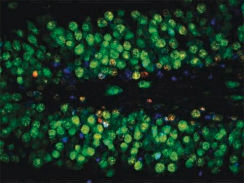
NeuN antibody (NEUronal Nuclei; clone A60) specifically recognizes the DNA-binding, neuron-specific protein NeuN, which is present in most CNS and PNS neuronal cell types of all vertebrates tested. NeuN protein distributions are apparently restricted to neuronal nuclei, perikarya and some proximal neuronal processes in both fetal and adult brain although, some neurons fail to be recognized by NeuN at all ages: INL retinal cells, Cajal-Retzius cells, Purkinje cells, inferior olivary and dentate nucleus neurons, and sympathetic ganglion cells are examples (Mullen et al., 1992; Wolf et al., 1996). Immunohistochemically detectable NeuN protein first appears at developmental timepoints that correspond with the withdrawal of the neuron from the cell cycle and/or with the initiation of terminal differentiation of the neuron (Mullen et al., 1992). Immunoreactivity appears around E9.5 in the mouse neural tube and is extensive throughout the developing nervous system by E12.5. Strong nuclear staining suggests a nuclear regulatory protein function; however, no evidence currently exists as to whether the NeuN protein antigen has a function in the distal cytoplasm or whether it is merely synthesized there before being transported back into the nucleus. No difference between protein isolated from purified nuclei and whole brain extract on immunoblots has been found (Mullen et al., 1992).

Specificity

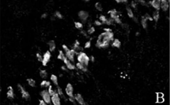
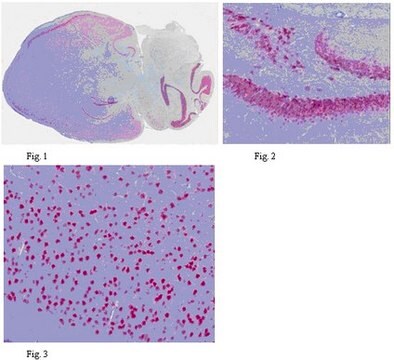
NeuN (or Neuronal Nuclei) reacts with most neuronal cell types throughout the nervous system of mice including cerebellum, cerebral cortex, hippocampus, thalamus, spinal cord and neurons in the peripheral nervous system including dorsal root ganglia, sympathetic chain ganglia and enteric ganglia.

Immunogen

Purified cell nuclei from mouse brain.

Application

Detect NeuN using this Anti-NeuN Antibody, clone A60, Cy3 Conjugate validated for use in IC.
Research Category
Neuroscience
Research Sub Category
Developmental Neuroscience

Quality

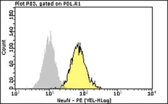
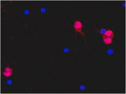
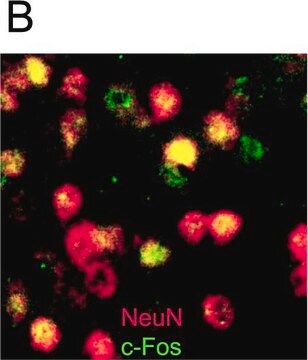
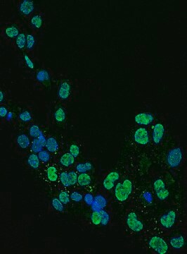
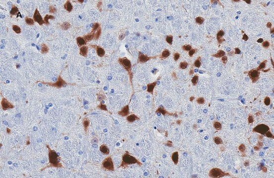
Evaluated by Immunocytochemistry in rat E18 primary cortex cells.

Immunocytochemistry Analysis: A 1:100 dilution of this antibody detected NeuN in rat E18 primary cortex cells.

Target description

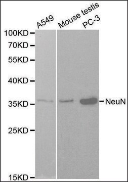
The unconjugated parent antibody (Cat. No. MAB377) has an observed molecular weight in the 46-48 kDa range and possibly another band at approximately 66 kDa

Physical form

Protein A purified
Purified mouse monoclonal IgG1 conjugated to Cy3 in PBS with 0.1% sodium azide and 15 mg/mL BSA.

Storage and Stability

Maintain refrigerated at 2-8 °C protected from light in undiluted aliquots for up to 6 months from date of receipt.

Analysis Note

Control
Rat E18 primary cortex neurons.

Other Notes

Unconjugated Anti-NeuN has been shown to work with multiple species including Human, Mouse, Ferret, Chicken, and Avian.

Disclaimer

Unless otherwise stated in our catalog or other company documentation accompanying the product(s), our products are intended for research use only and are not to be used for any other purpose, which includes but is not limited to, unauthorized commercial uses, in vitro diagnostic uses, ex vivo or in vivo therapeutic uses or any type of consumption or application to humans or animals.

Not finding the right product?  

Try our Product Selector Tool.

Storage Class

10 - Combustible liquids

wgk_germany

WGK 2

flash_point_f

Not applicable

flash_point_c

Not applicable


Certificates of Analysis (COA)

Search for Certificates of Analysis (COA) by entering the products Lot/Batch Number. Lot and Batch Numbers can be found on a product’s label following the words ‘Lot’ or ‘Batch’.

Already Own This Product?

Find documentation for the products that you have recently purchased in the Document Library.

Visit the Document Library

Judith Niesen et al.
iScience, 26(9), 107501-107501 (2023-08-23)
The zinc-finger transcription factor GLI2 is frequently amplified in childhood medulloblastoma of the Sonic-hedgehog type (SHH-MB), with or without amplification of NMYC or deletion of TP53. Despite the aggressive tumor behavior, tumorigenesis is not well understood, and adequate mouse models
Kyla Brown et al.
Human molecular genetics, 25(3), 558-570 (2015-12-10)
Rett syndrome is caused by mutations in the X-linked MECP2 gene, which encodes a chromosomal protein that binds to methylated DNA. Mouse models mirror the human disorder and therefore allow investigation of phenotypes at a molecular level. We describe an
Lucas F R Nascimento et al.
Diabetes, 65(3), 673-686 (2015-10-30)
Apoptosis of hypothalamic neurons is believed to play an important role in the development and perpetuation of obesity. Similar to the hippocampus, the hypothalamus presents constitutive and stimulated neurogenesis, suggesting that obesity-associated hypothalamic dysfunction can be repaired. Here, we explored
Larissa M G Cassiano et al.
Journal of neuroinflammation, 19(1), 272-272 (2022-11-16)
Thiamine (vitamin B1) is a cofactor for enzymes of central energy metabolism and its deficiency (TD) impairs oxidative phosphorylation, increases oxidative stress, and activates inflammatory processes that can lead to neurodegeneration. Wernicke-Korsakoff syndrome (WKS) is a consequence of chronic TD
Sangjune Kim et al.
Neuron, 103(4), 627-641 (2019-07-01)
Analysis of human pathology led Braak to postulate that α-synuclein (α-syn) pathology could spread from the gut to brain via the vagus nerve. Here, we test this postulate by assessing α-synucleinopathy in the brain in a novel gut-to-brain α-syn transmission

Our team of scientists has experience in all areas of research including Life Science, Material Science, Chemical Synthesis, Chromatography, Analytical and many others.

Contact Technical Service