Skip to Content
MilliporeSigma
All Photos(1)

Key Documents

CBL202

Sigma-Aldrich

Anti-Vimentin Antibody, clone VIM 3B4

clone VIM 3B4, Chemicon®, from mouse

Sign Into View Organizational & Contract Pricing


About This Item

UNSPSC Code:
12352203
eCl@ss:
32160702
NACRES:
NA.41

biological source

mouse

Quality Level

antibody form

purified immunoglobulin

antibody product type

primary antibodies

clone

VIM 3B4, monoclonal

species reactivity

chicken, amphibian, bovine, monkey, canine, human

manufacturer/tradename

Chemicon®

technique(s)

ELISA: suitable
immunofluorescence: suitable
immunohistochemistry: suitable (paraffin)
western blot: suitable

isotype

IgG2a

NCBI accession no.

UniProt accession no.

shipped in

wet ice

Gene Information

human ... VIM(7431)

Specificity

Highly specific for the intermediate filament protein vimentin which is present in all cells of mesenchymal origin. This antibody is positive with cells of mesenchymal derivation, including endothelial cells and certain smooth muscle cells of the vascular tract, fibroblasts, connective tissue, all types of blood cells, including thymocytes, interstitial and testicular Sertoli cells, ovarian follicle cells. It can also be used to detect coexpression of vimentin in combination with other intermediate filament proteins. VIM 3B4 has turned out to be the most avid monoclonal antibody to vimentin. Tumors specifically detected: sarcoma (including myosarcoma), lymphoma, melanoma. The binding region of monoclonal antibody VIM3B4 has been characterized by Bohn et al. (1992). According to these authors, the epitope has been localized on the alpha-helical part of vimentin (rod domain coil 2). Due to an aa substitution at position of aa 353 in murine vimentin (that could explain for the weak cross-reaction of the antibody with murine vimentin) they were able to narrow down the binding region around position 353. These findings were confirmed by truncation mutagenesis experiments using human vimentin (Rogers et al., 1995). Polypeptide reacting: MW 57 kDa intermediate filament protein (vimentin) of mesenchymal cells.

Immunogen

Vimentin purified from bovine lens

Application

Western blot
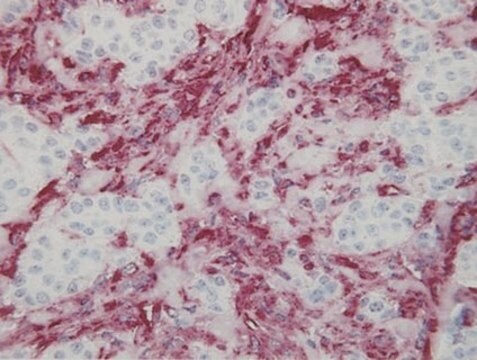
Immunohistochemistry: 1:100; Frozen and paraffin-embedded tissue sections with a minimum of a one hour incubation (longer for paraffin); protease pretreatment is recommended for paraffin-embedded sections.
Immunofluorescence
ELISA

Optimal working dilutions must be determined by the end user.
Research Category
Cell Structure
Research Sub Category
Cytoskeleton
This Anti-Vimentin Antibody, clone VIM 3B4 is validated for use in ELISA, IF, IH(P), WB for the detection of Vimentin.

Target description

57 kDa

Physical form

Format: Purified
Protein A Purified immunoglobulin presented as lyophilized material. Reconstitute with 1ml distilled water. Final solution is PBS, pH 7.4, containing 0.5% BSA and 0.09% sodium azide.
Protein A purified

Storage and Stability

Maintain at 2–8°C for up to 12 months from date of receipt.

Analysis Note

Control
Positive in RD cells, glioma cells, fibroblasts (SV-80), and MDCK cell line

Other Notes

Concentration: Please refer to the Certificate of Analysis for the lot-specific concentration.

Legal Information

CHEMICON is a registered trademark of Merck KGaA, Darmstadt, Germany

Disclaimer

Unless otherwise stated in our catalog or other company documentation accompanying the product(s), our products are intended for research use only and are not to be used for any other purpose, which includes but is not limited to, unauthorized commercial uses, in vitro diagnostic uses, ex vivo or in vivo therapeutic uses or any type of consumption or application to humans or animals.

Not finding the right product?  

Try our Product Selector Tool.

pictograms

Exclamation mark

signalword

Warning

Hazard Classifications

Acute Tox. 4 Dermal - Acute Tox. 4 Inhalation - Aquatic Chronic 3

Storage Class

11 - Combustible Solids

wgk_germany

WGK 3


Certificates of Analysis (COA)

Search for Certificates of Analysis (COA) by entering the products Lot/Batch Number. Lot and Batch Numbers can be found on a product’s label following the words ‘Lot’ or ‘Batch’.

Already Own This Product?

Find documentation for the products that you have recently purchased in the Document Library.

Visit the Document Library

Cytoskeletal components of lymphoid organs. I. Synthesis of cytokeratins 8 and 18 and desmin in subpopulations of extrafollicular reticulum cells of human lymph nodes, tonsils, and spleen.
Franke, W W and Moll, R
Differentiation, 36, 145-163 (1987)
Identification of differentially expressed genes in fetal rat forebrain exposed to a teratogen by cDNA microarray analysis.
S T Dheen,A J Hao,J Fu,P Gopalakrishnakone,E A Ling
Histology and Histopathology null
Cell type heterogeneity of intermediate filament expression in epithelia of the human pituitary gland.
Kasper, M, et al.
Histochemistry, 93, 93-103 (1989)
Lijie Liu et al.
Development, growth & differentiation, 49(8), 669-681 (2007-09-21)
To identify proteins involved in pancreatic development, we used a differential proteomics approach by comparing pancreatic extracts from four biologically significant stages of development: embryonic day (E) 15.5, E18.5, postnatal (P) days 0 and adult. By two-dimensional gel electrophoresis (2D-E)
Sites of origin and developmental dynamics of the neurons in the core and shell regions of torus semicircularis in the Chinese softshell turtle (Pelodiscus sinensis).
Chao Xi,Qiong Chen,Shao-Ju Zeng,Yu-Tao Lin,Yu-Fang Huang,Yu Liu,Xin-Wen Zhang,Ming-Xue Zuo
The Journal of Comparative Neurology null

Our team of scientists has experience in all areas of research including Life Science, Material Science, Chemical Synthesis, Chromatography, Analytical and many others.

Contact Technical Service