Skip to Content
MilliporeSigma
All Photos(3)

Key Documents

ABS2082

Sigma-Aldrich

Anti-PDH

serum, from rabbit

Synonym(s):

Pyruvate dehydrogenase E1 component subunit alpha, somatic form, mitochondrial, PDHE1-A type I, Pyruvate dehydrogenase E1 component subunit beta, mitochondrial, PDHE1-B

Sign Into View Organizational & Contract Pricing


About This Item

UNSPSC Code:
12352203
eCl@ss:
32160702
NACRES:
NA.41

biological source

rabbit

Quality Level

antibody form

serum

antibody product type

primary antibodies

clone

polyclonal

species reactivity

mouse, human

technique(s)

immunocytochemistry: suitable
immunofluorescence: suitable
immunohistochemistry: suitable
western blot: suitable

NCBI accession no.

UniProt accession no.

shipped in

dry ice

target post-translational modification

unmodified

Gene Information

human ... PDHA1(5160)

General description

Pyruvate dehydrogenase E1 component subunits alpha and beta, mitochondrial (EC 1.2.4.1; UniProt P08559 and P11177; also known as PDHE1-A type I and PDHE1-B, respectively) are encoded by the PDHA1 (also known as PDHA, PDHAD, PHE1A) and the PDHB (also known as PDHBD, PHE1B) genes (Gene ID 5160 and 5162, respectively) in human. The pyruvate dehydrogenase (PDH) complex catalyzes the overall, irreversible conversion of pyruvate to acetyl-CoA and CO2, and thereby links the glycolytic pathway to the tricarboxylic cycle. This complex contains multiple copies of three enzymatic components: PDH (E1 or PDHE1-A type I), dihydrolipoamide acetyltransferase (E2), and lipoamide dehydrogenase (E3). PDH (E1) performs the first two reactions within the PDH complex: a decarboxylation of pyruvate and a reductive acetylation of lipoic acid. Lipoic acid is covalently bound to dihydrolipoamide acetyltransferase (E2), which is the second catalytic component enzyme of PDC. The reaction catalyzed by PDH (E1) is considered to be the rate-limiting step for the PDH complex. PDH is regulated by both pyruvate dehydrogenase kinase (PDK)-mediated phosphorylation and feedback inhibition. Phosphorylation at Ser-232, Ser-293 and Ser-300 by PDK family kinases inactivates the enzyme; for this phosphorylation at a single site is sufficient. Dephosphorylation at all three sites, Ser-232, Ser-293, and Ser-300, is required for reactivation. PDH (E1) deficiency has been linked to primary lactic acidosis in children.

Specificity

This polyclonal antiserum detects both PDH alpha and beta subunits (Pliss, L., et al. (2004). Neurochem. 91(5):1082-1091).

Immunogen

Co-expressed recombinant full-length mature human PDH E1 alpha and beta subunits.

Application

Detect Pyruvate dehydrogenase E1 component subunit alpha, somatic form, mitochondrial using this rabbit polyclonal Anti-PDH, Cat. No. ABS2082, validated for use in Immunocytochemistry, Immunofluorescence, Immunohistochemistry, and Western Blotting.
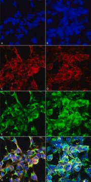
Immunocytochemistry Analysis: A 1:300 dilution from a representative lot detected PDH immunoreactivity in 4% paraformaldehyde-fixed, 1% Triton X-100-permeabilized BE(2)C human neuroblastoma cells (Courtesy of Professor Mulchand S. Patel, Ph.D., SUNY Buffalo, NY, U.S.A).

Western Blotting Analysis: A 1:4,000 dilution from a representative lot detected endogenous PDH alpha and beta subunits in 50 µg of mouse liver tissue lysate as well as 50 ng of purified recombinant human PDH alpha and beta subunits (Courtesy of Professor Mulchand S. Patel, Ph.D., SUNY Buffalo, NY, U.S.A).

Immunofluorescence Analysis: A representative lot immunostained oocytes in Bouins fluid-fixed, paraffin-embedded sections of mouse ovaries. A loss of oocyte PDH immunostaining was observed in ovaries from transgenic mice carrying homozygous floxed Pdha1 alleles and zona pellucida protein-3 (Zp3) promoter-driven Cre expression to allow oocyte-specific Pdha1 deletion (Johnson, M.T., et al. (2007). Biol Reprod. 77(1):2-8)

Immunohistochemistry Analysis: A representative lot immunostained neural cells in paraplast-embedded brain microtome sections from Bouins fluid-perfused mice. A marked increase in cells with low PDH immunostaining was observed in brain from transgenic mice carrying heterozygous floxed Pdha1 allele and nestin promoter-driven Cre expression to allow brain tissue-specific Pdha1 deletion (Pliss, L., et al. (2004). Neurochem. 91(5):1082-1091).

Western Blotting Analysis: A representative lot detected reduced level of PDH alpha & beta subunits in brain, but not liver, tissue from transgenic mice carrying heterozygous floxed Pdha1 allele and nestin promoter-driven Cre expression to allow brain tissue-specific Pdha1 deletion (Pliss, L., et al. (2004). Neurochem. 91(5):1082-1091).
Research Category
Signaling

Quality

Evaluated by Western Blotting in human brain.

Western Blotting Analysis: A 1:2,000 dilution of this antibody detected PDH alpha and beta subunits in 10 µg of human whole brain tissue lysate.

Target description

~43/37 kDa observed. 40.08/35.90 kDa (human alpha/beta subunit; transit peptide removed; UniProt P08559/P11177), 40.18/35.77 kDa (mouse alpha/beta subunit; transit peptide removed; UniProt P35486/Q9D051) Uncharacterized bands may be observed in some lysate(s).

Physical form

Rabbit polyclonal antibody serum with 0.05% sodium azide.
Unpurified.

Storage and Stability

Stable for 1 year at -20°C from date of receipt.

Handling Recommendations: Upon receipt and prior to removing the cap, centrifuge the vial and gently mix the solution. Aliquot into microcentrifuge tubes and store at -20°C. Avoid repeated freeze/thaw cycles, which may damage IgG and affect product performance.

Other Notes

Concentration: Please refer to lot specific datasheet.

Disclaimer

Unless otherwise stated in our catalog or other company documentation accompanying the product(s), our products are intended for research use only and are not to be used for any other purpose, which includes but is not limited to, unauthorized commercial uses, in vitro diagnostic uses, ex vivo or in vivo therapeutic uses or any type of consumption or application to humans or animals.

Not finding the right product?  

Try our Product Selector Tool.

Storage Class

12 - Non Combustible Liquids

wgk_germany

WGK 1


Certificates of Analysis (COA)

Search for Certificates of Analysis (COA) by entering the products Lot/Batch Number. Lot and Batch Numbers can be found on a product’s label following the words ‘Lot’ or ‘Batch’.

Already Own This Product?

Find documentation for the products that you have recently purchased in the Document Library.

Visit the Document Library

Joongyu D Song et al.
Cell metabolism, 32(5), 726-735 (2020-10-10)
Alterations in muscle mitochondrial substrate preference have been postulated to play a major role in the pathogenesis of muscle insulin resistance. In order to examine this hypothesis, we assessed the ratio of mitochondrial pyruvate oxidation (VPDH) to rates of mitochondrial
Charandeep Singh et al.
Nature communications, 11(1), 1277-1277 (2020-03-11)
Although supplemental oxygen is required to promote survival of severely premature infants, hyperoxia is simultaneously harmful to premature developing tissues such as in the retina. Here we report the effect of hyperoxia on central carbon metabolism in primary mouse Müller
Graeme A M Nimmo et al.
Human molecular genetics, 28(2), 290-306 (2018-10-12)
LonP1 is crucial for maintaining mitochondrial proteostasis and mitigating cell stress. We identified a novel homozygous missense LONP1 variant, c.2282 C > T, (p.Pro761Leu), by whole-exome and Sanger sequencing in two siblings born to healthy consanguineous parents. Both siblings presented with stepwise
Chantal R Ryan et al.
Nutrients, 13(7) (2021-08-11)
White adipose tissue (WAT) is a dynamic endocrine organ that can play a significant role in thermoregulation. WAT has the capacity to adopt structural and functional characteristics of the more metabolically active brown adipose tissue (BAT) and contribute to non-shivering

Our team of scientists has experience in all areas of research including Life Science, Material Science, Chemical Synthesis, Chromatography, Analytical and many others.

Contact Technical Service