Skip to Content
MilliporeSigma
All Photos(2)

Key Documents

ABN182

Sigma-Aldrich

Anti-CYP27B1 Antibody

from rabbit, purified by affinity chromatography

Synonym(s):

25-hydroxyvitamin D-1 alpha hydroxylase, mitochondrial, 25-OHD-1 alpha-hydroxylase, 25-hydroxyvitamin D(3) 1-alpha-hydroxylase, VD3 1A hydroxylase, Calcidiol 1-monooxygenase, Cytochrome P450 subfamily XXVIIB polypeptide 1, Cytochrome P450C1 alpha, Cytoch

Sign Into View Organizational & Contract Pricing


About This Item

UNSPSC Code:
12352203
eCl@ss:
32160702
NACRES:
NA.41

biological source

rabbit

Quality Level

antibody form

affinity isolated antibody

antibody product type

primary antibodies

clone

polyclonal

purified by

affinity chromatography

species reactivity

mouse, rat, human

species reactivity (predicted by homology)

chimpanzee (based on 100% sequence homology)

technique(s)

immunohistochemistry: suitable
western blot: suitable

NCBI accession no.

UniProt accession no.

shipped in

wet ice

target post-translational modification

unmodified

Gene Information

human ... CYP27B1(1594)

General description

25-hydroxyvitamin D-1 alpha hydroxylase (1αOHase; VD3 1A hydroxylase; 25-OHD-1 alpha-hydroxylase; 25-hydroxyvitamin D(3) 1-alpha-hydroxylase; or CYP27B1) is a mitochondrial enzyme that belongs to the cytochrome P450 family. It catalyzes the hydroxylation of 25-hydroxyvitamin D3 to form an active 1-alpha, 25-dihydroxyvitamin D3, in the kidneys and other tissues. 25-hydroxyvitamin D-1 alpha hydroxylase plays an important role in bone growth, calcium metabolism, and tissue differentiation. Previous studies have reported that mice deficient in 25-hydroxyvitamin D-1 alpha hydroxylase are affected by hypocalcemia, secondary hyperparathyroidism, retarded growth, and the skeletal abnormalities. In humans, defects in this enzyme causes pseudovitamin D deficiency rickets (vitamin D-dependent type 1A rickets), an autosomal recessive condition characterized by abnormal bone mineralization and impaired growth.

Immunogen

KLH-conjugated linear peptide corresponding to human CYP27B1.

Application

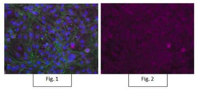
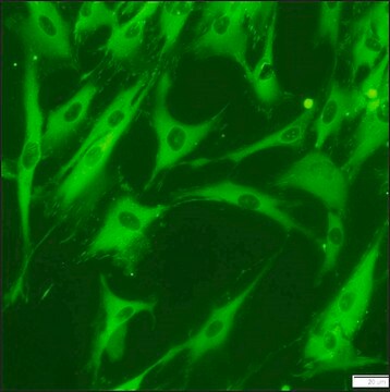
Immunohistochemistry Analysis: A 1:1,000 dilution from a representative lot detected CYP27B1 in mouse and rat hippocampus tissue and in rat kidney tissue.
Research Category
Neuroscience
Research Sub Category
Developmental Neuroscience
This Anti-CYP27B1 Antibody is validated for use in Western Blotting, IHC for the detection of CYP27B1.

Quality

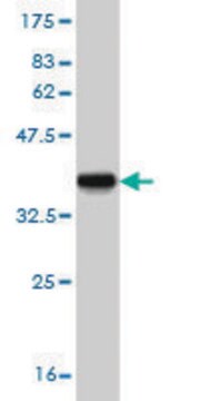
Evaluated by Western Blot in serum free MCF-7 cell lysate.

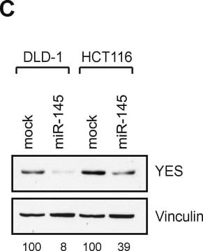
Western Blot Analysis: 1 µg/mL of this antibody detected CYP27B1 in 10 µg of serum free MCF-7 cell lysate.

Target description

~58 kDa observed. Uncharacterized bands may be observed at ~55 kDa, ~70 kDa, ~110 kDa, and ~115 kDa in some cell lysates. A doublet at ~58 kDa may be observed in some cell lysates (Kemmis, C. M., et al. (2006). J Nutr. 136(4):887-892.).

Physical form

Affinity purified
Purified rabbit polyclonal in buffer containing 0.1 M Tris-Glycine (pH 7.4), 150 mM NaCl with 0.05% sodium azide.

Storage and Stability

Stable for 1 year at 2-8°C from date of receipt.

Analysis Note

Control
Serum free MCF-7 cell lysate

Disclaimer

Unless otherwise stated in our catalog or other company documentation accompanying the product(s), our products are intended for research use only and are not to be used for any other purpose, which includes but is not limited to, unauthorized commercial uses, in vitro diagnostic uses, ex vivo or in vivo therapeutic uses or any type of consumption or application to humans or animals.

Not finding the right product?  

Try our Product Selector Tool.

Storage Class

12 - Non Combustible Liquids

wgk_germany

WGK 1

flash_point_f

Not applicable

flash_point_c

Not applicable


Certificates of Analysis (COA)

Search for Certificates of Analysis (COA) by entering the products Lot/Batch Number. Lot and Batch Numbers can be found on a product’s label following the words ‘Lot’ or ‘Batch’.

Already Own This Product?

Find documentation for the products that you have recently purchased in the Document Library.

Visit the Document Library

Eva S Liu et al.
Endocrinology, 155(10), 3750-3756 (2014-07-25)
Phosphate and parathyroid hormone related peptide (PTHrP) are required for normal growth plate maturation. Hypophosphatemia impairs hypertrophic chondrocyte apoptosis leading to rachitic expansion of the growth plate; however, the effect of phosphate restriction on chondrocyte differentiation during endochondral bone formation
Xiaochen Li et al.
Frontiers in pharmacology, 12, 690181-690181 (2021-06-22)
Osteoarthritis (OA) is a worldwide degenerative joint disease that seriously impaired the quality of life of patients. OA has been established as a disease with metabolic disorder. Cholesterol 25-hydroxylase (CH25H) was proved to play a key role in cartilage cholesterol
Hiroki Maruyama et al.
FASEB bioAdvances, 2(6), 365-381 (2020-07-04)
Low bone mineral density (BMD)-diagnosed as osteoporosis or osteopenia-has been reported as a new characteristic feature of Fabry disease; however, the mechanism underlying the development of low BMD is unknown. We previously revealed that a mouse model of Fabry disease
Samanta Mazzetti et al.
CNS neuroscience & therapeutics, 28(5), 703-713 (2022-02-16)
Astrocytes are involved in Parkinson's disease (PD) where they could contribute to α-Synuclein pathology but also to neuroprotection via α-Synuclein clearance. The molecular signature underlying their dual role is still elusive. Given that vitamin D has been recently suggested to

Our team of scientists has experience in all areas of research including Life Science, Material Science, Chemical Synthesis, Chromatography, Analytical and many others.

Contact Technical Service