Skip to Content
MilliporeSigma
All Photos(8)

Documents

AB5804

Sigma-Aldrich

Anti-Glial Fibrillary Acidic Protein (GFAP) Antibody

serum, Chemicon®

Synonym(s):

glial fibrillary acidic protein

Sign Into View Organizational & Contract Pricing


About This Item

UNSPSC Code:
12352203
eCl@ss:
32160702
NACRES:
NA.41

biological source

rabbit

Quality Level

antibody form

serum

antibody product type

primary antibodies

clone

polyclonal

species reactivity

human, bovine, canine, rat

manufacturer/tradename

Chemicon®

technique(s)

immunocytochemistry: suitable
immunohistochemistry (formalin-fixed, paraffin-embedded sections): suitable
western blot: suitable

NCBI accession no.

UniProt accession no.

shipped in

dry ice

target post-translational modification

unmodified

Gene Information

human ... GFAP(2670)

General description

Glial fibrillary acidic protein is a class-III intermediate filament. GFAP is the main constituent of intermediate filaments in astrocytes and serves as a cell specific marker that distinguishes differentiated astrocytes from other glial cells during the development of the central nervous system.

Specificity

It is expected that the antibody will react with most mammalian GFAP due to homology. Reactivity with other species has not been confirmed.
Recognizes Glial Fibrillary Acidic Protein (GFAP).

Immunogen

Purified bovine GFAP

Application

Anti-Glial Fibrillary Acidic Protein (GFAP) Antibody is an antibody against Glial Fibrillary Acidic Protein (GFAP) for use in IC, IH, IH(P) with more than 65 product citations.
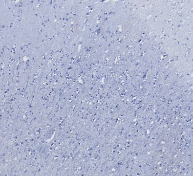
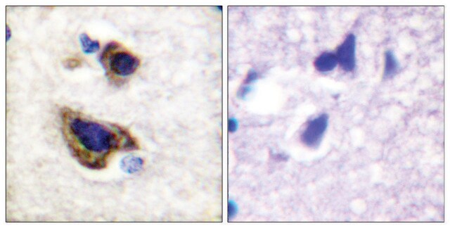
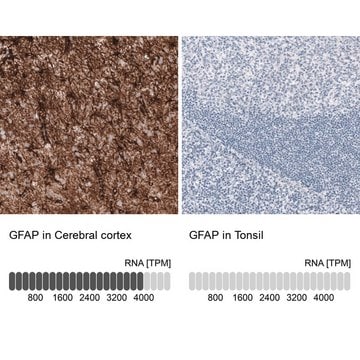
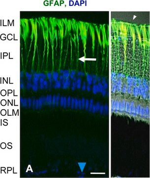
Immunohistochemistry(paraffin):
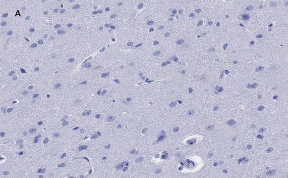
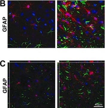
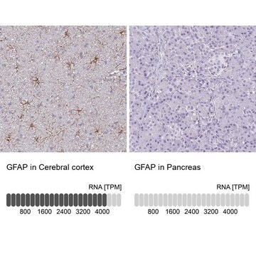
Representative images from a previous lot.

Immunocytochemistry:
A 1:1,000 dilution of a previous lot was used in Immunocytochemistry.
Research Category
Neuroscience
Research Sub Category
Neuronal & Glial Markers

Quality

Routinely evaluated by immunohistochemistry on brain tissue, astrocytes, and neurons.

Immunohistochemistry(paraffin) Analysis:
GFAP (IHC2078) staining of Human Brain. Tissue pretreated with Citrate Buffer, pH 6.0. Pre-diluted polyclonal antibody, using IHC-Select Detection with HRP-DAB. Glial cells stain strongly (brown).
Optimal Staining with Citrate Buffer Epitope Retrieval: Human Brain

Target description

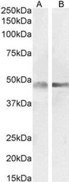
50 kDa

Linkage

Replaces: AB1540

Physical form

Rabbit serum. Liquid. Contains no preservative.
Unpurified

Storage and Stability

Stable for 1 year at -20°C undiluted aliquots from date of receipt.
Handling Recommendations: Upon receipt, and prior to removing the cap, centrifuge the vial and gently mix the solution. Aliquot into microcentrifuge tubes and store at -20°C. Avoid repeated freeze/thaw cycles, which may damage IgG and affect product performance.

Analysis Note

Control
Mouse brain tissue, rat astrocytes, rat neurons.

Legal Information

CHEMICON is a registered trademark of Merck KGaA, Darmstadt, Germany

Disclaimer

Unless otherwise stated in our catalog or other company documentation accompanying the product(s), our products are intended for research use only and are not to be used for any other purpose, which includes but is not limited to, unauthorized commercial uses, in vitro diagnostic uses, ex vivo or in vivo therapeutic uses or any type of consumption or application to humans or animals.

Not finding the right product?  

Try our Product Selector Tool.

Storage Class

10 - Combustible liquids

wgk_germany

WGK 1

flash_point_f

Not applicable

flash_point_c

Not applicable


Certificates of Analysis (COA)

Search for Certificates of Analysis (COA) by entering the products Lot/Batch Number. Lot and Batch Numbers can be found on a product’s label following the words ‘Lot’ or ‘Batch’.

Already Own This Product?

Find documentation for the products that you have recently purchased in the Document Library.

Visit the Document Library

Damir Sapunar et al.
Pain, 152(5), 1173-1181 (2011-03-08)
Neuropeptide Y (NPY) has an important but still insufficiently defined role in pain modulation. We therefore examined the ability of NPY to modulate experimentally induced neuropathic pain by injecting it directly into dorsal root ganglion (DRG) immediately following spinal nerve
Inhibition of CXCR1 and CXCR2 chemokine receptors attenuates acute inflammation, preserves gray matter and diminishes autonomic dysreflexia after spinal cord injury.
Marsh DR, Flemming JM
Spinal Cord null
Astrogliosis in EAE spinal cord: derivation from radial glia, and relationships to oligodendroglia.
Peter Bannerman,Ashleigh Hahn,Athena Soulika,Vittorio Gallo,David Pleasure
Glia null
Congenital anomalies and rhabdoid tumor associated with 22q11 germline deletion and somatic inactivation of the SMARCB1 tumor suppressor.
Toth, G; Zraly, CB; Thomson, TL; Jones, C; Lapetino, S; Muraskas, J; Zhang, J; Dingwall, AK
Genes Chromosomes Cancer null
Expression patterns and subcellular localization of the Down syndrome candidate protein MNB/DYRK1A suggest a role in late neuronal differentiation
Hammerle, B. et al.
The European Journal of Neuroscience, 17(11), 2277-2286 (2003)

Articles

ReNcell neural progenitors are immortalized human neural stem cell lines that can differentiate into neurons, astrocytes sand oligodendrocytes.

Our team of scientists has experience in all areas of research including Life Science, Material Science, Chemical Synthesis, Chromatography, Analytical and many others.

Contact Technical Service