Przejdź do zawartości
Merck
Wszystkie zdjęcia(1)

Kluczowe dokumenty

MABF261

Sigma-Aldrich

Anti-PD-1 Antibody, clone 16A2.1

clone 16A2.1, from mouse, purified by affinity chromatography

Synonim(y):

PD-1, Programmed cell death protein 1, Protein PD-1, hPD-1, CD279

Zaloguj sięWyświetlanie cen organizacyjnych i kontraktowych


About This Item

Kod UNSPSC:
12352203
eCl@ss:
32160702
NACRES:
NA.41

pochodzenie biologiczne

mouse

Poziom jakości

forma przeciwciała

purified immunoglobulin

rodzaj przeciwciała

primary antibodies

klon

16A2.1, monoclonal

oczyszczone przez

affinity chromatography

reaktywność gatunkowa

human

metody

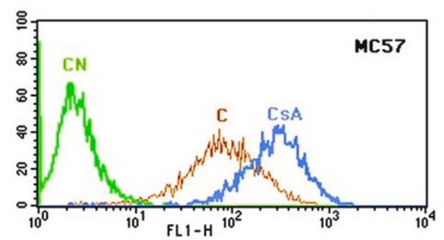
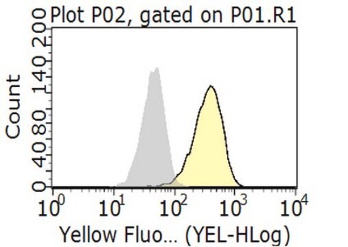
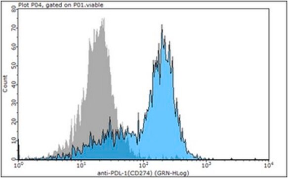
flow cytometry: suitable
western blot: suitable

izotyp

IgG2aκ

numer dostępu NCBI

numer dostępu UniProt

Warunki transportu

wet ice

docelowa modyfikacja potranslacyjna

unmodified

informacje o genach

human ... PDCD1(5133)

Opis ogólny

Programmed cell death protein 1 (PD-1) (UniProt Q15116), also known as CD279, hPD-1, hPD-I, hSLE1, Protein PD-1, Systemic lupus erythematosus susceptibility 2) is a type I single transmembrane protein encoded by the PDCD1 gene in human. Maintenance of peripheral tolerance by preventing the development of autoimmunity due to T lymphocytes specific for self-antigens is essential for homeostasis of the immune system. PD-1 receptor CD279 and its ligands PD-L1 (B7-H1, CD274) and PD-L2 (B7-DC; CD273) play a vital role in the induction and maintenance of peripheral tolerance. Upregulated PD-1 expression is seen on conventional CD4+ CD25- T cells (Tconv) when exposed to mesenchymal stem cells, resulting in increased Tconv apoptosis due to PD-1 stimulation by the MSC PD-L2.

Immunogen

Epitope: Extracellular domain
KLH-conjugated linear peptide corresponding to the extracellular domain of human PD-1.

Zastosowanie

Anti-PD-1 Antibody, clone 16A2.1 is an antibody against PD-1 for use in Western Blotting, Flow Cytometry.
Flow Cytometry Analysis: 2.0 µg of this antibody from a representative lot detected PD-1 in 1X10E6 Staph enterotoxin B treated human peripheral blood mononuclear cells.
Research Category
Inflammation & Immunology
Research Sub Category
Immunoglobulins & Immunology

Jakość

Evaluated by Western Blotting in TPA/LPS treated THP-1 cell lysate.

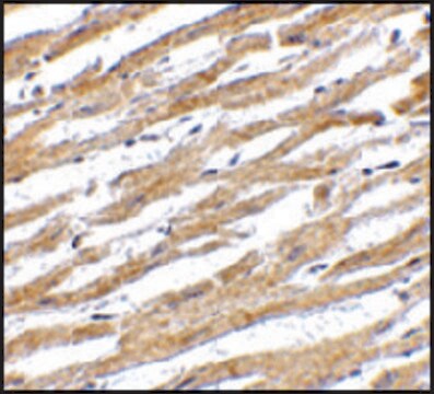
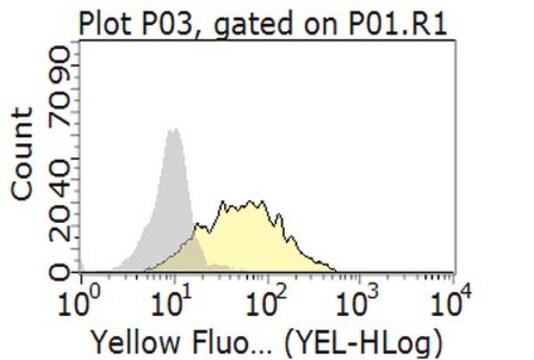
Western Blotting Analysis: 1.0 µg/mL of this antibody detected PD-1 in 10 µg of TPA/LPS treated THP-1 cell lysate.

Opis wartości docelowych

~32 kDa observed

Postać fizyczna

Protein G Purified
Purified mouse monoclonal IgG2ak in buffer containing 0.1 M Tris-Glycine (pH 7.4), 150 mM NaCl with 0.05% sodium azide.

Przechowywanie i stabilność

Stable for 1 year at 2-8°C from date of receipt.

Inne uwagi

Concentration: Please refer to lot specific datasheet.

Oświadczenie o zrzeczeniu się odpowiedzialności

Unless otherwise stated in our catalog or other company documentation accompanying the product(s), our products are intended for research use only and are not to be used for any other purpose, which includes but is not limited to, unauthorized commercial uses, in vitro diagnostic uses, ex vivo or in vivo therapeutic uses or any type of consumption or application to humans or animals.
Ta strona może zawierać tekst przetłumaczony maszynowo.

Nie możesz znaleźć właściwego produktu?  

Wypróbuj nasz Narzędzie selektora produktów.

Kod klasy składowania

12 - Non Combustible Liquids

Klasa zagrożenia wodnego (WGK)

WGK 1

Temperatura zapłonu (°F)

Not applicable

Temperatura zapłonu (°C)

Not applicable


Certyfikaty analizy (CoA)

Poszukaj Certyfikaty analizy (CoA), wpisując numer partii/serii produktów. Numery serii i partii można znaleźć na etykiecie produktu po słowach „seria” lub „partia”.

Masz już ten produkt?

Dokumenty związane z niedawno zakupionymi produktami zostały zamieszczone w Bibliotece dokumentów.

Odwiedź Bibliotekę dokumentów

Nasz zespół naukowców ma doświadczenie we wszystkich obszarach badań, w tym w naukach przyrodniczych, materiałoznawstwie, syntezie chemicznej, chromatografii, analityce i wielu innych dziedzinach.

Skontaktuj się z zespołem ds. pomocy technicznej