콘텐츠로 건너뛰기
Merck
모든 사진(1)

주요 문서

PKH26PCL

Sigma-Aldrich

PKH26 Red Fluorescent Cell Linker Kit for Phagocytic Cell Labeling

Distributed for Phanos Technologies

동의어(들):

Phagocytic cell label

로그인조직 및 계약 가격 보기


About This Item

UNSPSC 코드:
12352207
NACRES:
NA.32

애플리케이션

This kit is for phagocytic cell labeling. It is used to selectively label cells with phagocytic capabilities such as monocytes, macrophages or neutrophils.

원리

The labeling occurs through the formation of dye aggregates or particulates. The aggregate formation significantly inhibits the uptake of dye by non-phagocytic cells, such as lymphocytes, but facilitates dye uptake by phagocytic cells. Labeled cells appear patchy or spotted because the dye is localized in phagocytic compartments of the cells. The dye appears to be resistant to metabolic attack and has been found to remain with the cells for at least 21 days in vivo.
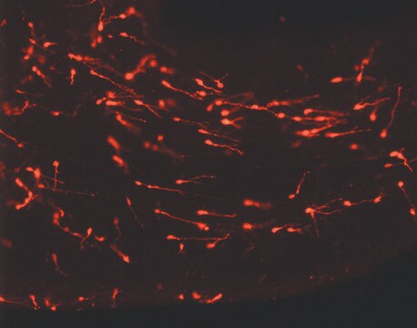
Labeling of phagocytic cells by this methodology may be conducted either in vitro or in vivo. Intraperitoneal or intravenous injections of the PKH26 labeling solution will successfully label phagocytic cells in vivo, while cells of interest which have been isolated may be stained using in vitro labeling methods.

결합

For additional technical details on PKH and CellVue® Fluorescent Cell Linker Dyes including an extensive bibliography, please visit here.

법적 정보

CellVue is a registered trademark of Phanos Technologies

키트 구성품 전용

제품 번호
설명

  • Diluent B 6 x 10

  • PKH26 cell linker in ethanol .5 mL

관련 제품

픽토그램

FlameExclamation mark

신호어

Danger

유해 및 위험 성명서

Hazard Classifications

Eye Irrit. 2 - Flam. Liq. 2

Storage Class Code

3 - Flammable liquids

Flash Point (°F)

57.2 °F - closed cup

Flash Point (°C)

14.0 °C - closed cup


가장 최신 버전 중 하나를 선택하세요:

시험 성적서(COA)

Lot/Batch Number

적합한 버전을 찾을 수 없으신가요?

특정 버전이 필요한 경우 로트 번호나 배치 번호로 특정 인증서를 찾을 수 있습니다.

이 제품을 이미 가지고 계십니까?

문서 라이브러리에서 최근에 구매한 제품에 대한 문서를 찾아보세요.

문서 라이브러리 방문

Hyung-Syup Kim et al.
Journal of Korean Neurosurgical Society, 44(4), 249-255 (2008-12-20)
In Moyamoya disease, the primary goal of treatment is to improve collateral circulation through angiogenesis. In the present study, we obtained and sub-cultured bone marrow stromal cells (BMSCs) from rats without a cell-mediated immune response. Then, we injected the labeled
Jaime Murphy et al.
American journal of respiratory cell and molecular biology, 38(4), 380-385 (2008-01-15)
To further examine the half-life of alveolar macrophages, chimeric CD 45.2 mice were generated through bone marrow transplantation of donor CD 45.1 cells. Before administration of donor cells, recipient mice were divided into two cohorts: the first cohort received total
K Tabata et al.
Gene therapy, 18(10), 969-978 (2011-04-23)
We previously identified the mouse and human Glipr1 and GLIPR1/RTVP-1 genes, respectively, as direct p53 targets with proapoptotic activities in various cancer cell lines, including prostate cancer (PCa). Intratumoral injection of an adenoviral vector capable of efficient transduction and expression
Lara Campana et al.
Journal of immunology (Baltimore, Md. : 1950), 200(3), 1169-1187 (2017-12-22)
The disposal of apoptotic bodies by professional phagocytes is crucial to effective inflammation resolution. Our ability to improve the disposal of apoptotic bodies by professional phagocytes is impaired by a limited understanding of the molecular mechanisms that regulate the engulfment
Meaghan M Hunter et al.
Gastroenterology, 138(4), 1395-1405 (2010-01-05)
Infection with the rat tapeworm Hymenolepis diminuta reduces the severity of dinitrobenzene sulfonic acid (DNBS)-induced colitis in mice. Infection with H. diminuta increases colonic expression of arginase-1 and found in inflammatory zone 1 (FIZZ1), markers of alternatively activated macrophages (AAMs).

문서

PKH dyes are easy to use and achieve stable, uniform, and reproducible fluorescent labeling of live cells. PKH dyes are non-toxic membrane stains which produce high signal to noise ratio.

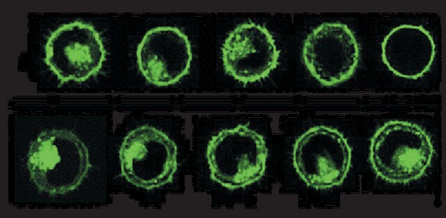
Lipophilic cell tracking dyes enable cancer biologists to track tumor and immune cell functions both in vitro and in vivo. Read the article to choose a right membrane dye kit for cell tracking and proliferation monitoring.

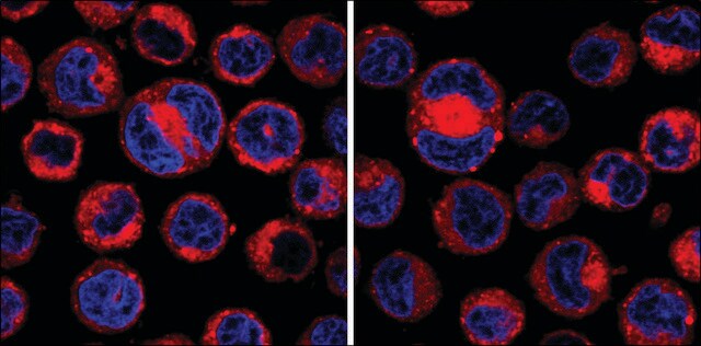
Optimal staining is a key component for studying tumorigenesis and progression. Learn useful tips and techniques for dye applications, including examples from recent studies.

A video about how you can use fluorescent cell tracking dyes in combination with flow and image cytometry to study interactions and fates of different cell types in vitro and in vivo.

모두 보기

자사의 과학자팀은 생명 과학, 재료 과학, 화학 합성, 크로마토그래피, 분석 및 기타 많은 영역을 포함한 모든 과학 분야에 경험이 있습니다..

고객지원팀으로 연락바랍니다.