콘텐츠로 건너뛰기
Merck
모든 사진(2)

Key Documents

PKH67GL

Sigma-Aldrich

PKH67 Green Fluorescent Cell Linker Kit for General Cell Membrane Labeling

Distributed for Phanos Technologies

동의어(들):

Green PKH membrane labeling kit

로그인조직 및 계약 가격 보기


About This Item

UNSPSC 코드:
12352207
NACRES:
NA.32

Quality Level

포장

pkg of 1 kit

저장 조건

protect from light

형광

λex 490 nm; λem 502 nm (PKH67 dye)

검출 방법

fluorometric

배송 상태

ambient

저장 온도

room temp

애플리케이션

PKH67 Green Fluorescent Cell Linker Kit for General Cell Membrane Labeling has been used:
  • To label and further investigate the exosomes expelled from cells in triple-negative breast cancer condition.
  • To label apoptotic cells, in order to study the role of ανβ5 receptor in both binding and internalization of apoptotic cells.
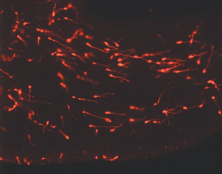
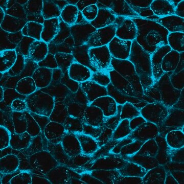
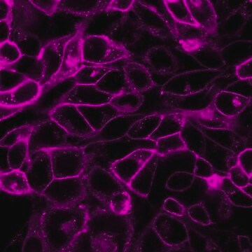
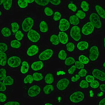
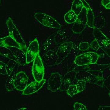
  • In fluorescence imaging.

This kit is for general cell membrane labeling. PKH67 has a longer aliphatic carbon tail than PKH1 and PKH2, two other green dyes previously described for in vitro and in vivo cell tracking. Based on the longer tail length, in-house studies have consistently shown reduced cell-cell transfer for PKH67 as compared to PKH2.
Slow loss of fluorescence has been observed in in vivo studies using PKH1 and PKH2. PKH67 may exhibit similar properties since this behavior appears to be characteristic of green cell linker dyes, but not red cell linker dyes. Correlation of in vitro cell membrane retention with in vivo fluorescence half life in non-dividing cells predicts an in vivo fluorescence half life of 10-12 days for PKH67. Other green cell linker dyes with similar half lives have been used to monitor in vivo lymphocyte and macrophage trafficking over periods of 1-2 months, suggesting that PKH67 will also be useful for in vivo tracking studies of moderate length.

결합

For additional technical details on PKH and CellVue® Fluorescent Cell Linker Dyes including an extensive bibliography, please visit here.

법적 정보

CellVue is a registered trademark of Phanos Technologies

키트 구성품 전용

제품 번호
설명

  • Diluent C 6 x 10

  • PKH67 Cell Linker in ethanol .5 mL

픽토그램

FlameExclamation mark

신호어

Danger

유해 및 위험 성명서

Hazard Classifications

Eye Irrit. 2 - Flam. Liq. 2

Storage Class Code

3 - Flammable liquids

Flash Point (°F)

57.2 °F - closed cup

Flash Point (°C)

14 °C - closed cup


시험 성적서(COA)

제품의 로트/배치 번호를 입력하여 시험 성적서(COA)을 검색하십시오. 로트 및 배치 번호는 제품 라벨에 있는 ‘로트’ 또는 ‘배치’라는 용어 뒤에서 찾을 수 있습니다.

이 제품을 이미 가지고 계십니까?

문서 라이브러리에서 최근에 구매한 제품에 대한 문서를 찾아보세요.

문서 라이브러리 방문

이미 열람한 고객

alpha v beta 5 integrin recruits the CrkII-Dock180-Rac1 complex for phagocytosis of apoptotic cells
Albert, Matthew L and Kim, Jong-Ii and Birge, Raymond B
Nature Cell Biology, 2(12), 899-899 (2000)
Increased expression of inducible HSP70 in apoptotic cells is correlated with their efficacy for antitumor vaccine therapy
Masse D, et al.
International Journal of Cancer. Journal International Du Cancer, 111(4), 575-583 (2004)
Exosomes from triple-negative breast cancer cells can transfer phenotypic traits representing their cells of origin to secondary cells
O Brien K, et al.
European Journal of Cancer, 49(8), 1845-1859 (2013)
Anjali P Kusumbe et al.
Stem cells (Dayton, Ohio), 27(3), 498-508 (2009-03-04)
Recruitment and localization of endothelial precursors within tumors is a potential area for the development of therapeutics, because their functional contribution to tumor vasculature is realized to be important for cancer cell survival. However, the exact nature of the recruited
Z M Wu et al.
Placenta, 33(3), 188-194 (2012-01-04)
As human blastocyst-derived extravillous trophoblasts (EVTs) invade the early decidua, they are positioned to interact with immune cells and resident decidual cells, and remodel spiral arteries into high capacity vessels that increase blood flow to the developing fetal-placental unit. Shallow

문서

PKH dyes are easy to use and achieve stable, uniform, and reproducible fluorescent labeling of live cells. PKH dyes are non-toxic membrane stains which produce high signal to noise ratio.

Lipophilic cell tracking dyes enable cancer biologists to track tumor and immune cell functions both in vitro and in vivo. Read the article to choose a right membrane dye kit for cell tracking and proliferation monitoring.

Optimal staining is a key component for studying tumorigenesis and progression. Learn useful tips and techniques for dye applications, including examples from recent studies.

A video about how you can use fluorescent cell tracking dyes in combination with flow and image cytometry to study interactions and fates of different cell types in vitro and in vivo.

모두 보기

자사의 과학자팀은 생명 과학, 재료 과학, 화학 합성, 크로마토그래피, 분석 및 기타 많은 영역을 포함한 모든 과학 분야에 경험이 있습니다..

고객지원팀으로 연락바랍니다.