콘텐츠로 건너뛰기
Merck
모든 사진(1)

주요 문서

251R-1

Sigma-Aldrich

Factor XIIIa (EP3372) Rabbit Monoclonal Primary Antibody

로그인조직 및 계약 가격 보기


About This Item

UNSPSC 코드:
12352203
NACRES:
NA.41

생물학적 소스

rabbit

Quality Level

100
500

결합

unconjugated

항체 형태

culture supernatant

항체 생산 유형

primary antibodies

클론

EP3372, monoclonal

설명

For In Vitro Diagnostic Use in Select Regions (See Chart)

양식

buffered aqueous solution

종 반응성

human

포장

vial of 0.1 mL concentrate (251R-14)
vial of 0.5 mL concentrate (251R-15)
bottle of 1.0 mL predilute (251R-17)
vial of 1.0 mL concentrate (251R-16)
bottle of 7.0 mL predilute (251R-18)

제조업체/상표

Cell Marque

기술

immunohistochemistry (formalin-fixed, paraffin-embedded sections): 1:100-1:500

동형

IgG

제어

dermatofibroma

배송 상태

wet ice

저장 온도

2-8°C

시각화

cytoplasmic

유전자 정보

human ... F13A1(2162)

일반 설명

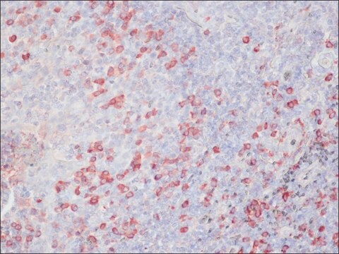
Factor XIIIa is a plasma transglutaminase that is part of the blood coagulation cascade and plays a critical role in stabilizing fibrin networks at the site of an injury to produce a clot. Factor XIIIa is expressed in platelets, megakaryocytes, fibroblast-like cells in the placenta, uterus, and prostate, monocytes and macrophages, and dermal dendritic cells. Anti-Factor XIIIa immunohistochemistry is used to identify fibrohistiocytic neoplasms such as dermatofibroma.
Factor XIIIa is a blood proenzyme that has been identified in platelets, megakaryocyte, and fibroblast-like mesenchymal or histiocytic cells present in the placenta, uterus, and prostate; it is also present in monocytes and macrophages and dermal dendritic cells. Anti- Factor XIIIa has been found to be useful in differentiating between dermatofibroma (90% (+)), dermatofibrosarcoma protuberans (25%(+)) and desmoplastic malignant melanoma (0%(+)). Factor XIIIa positivity is also seen in capillary hemagioblastoma (100%(+)), hemangioendothelioma (100%(+)), hemangiopericytoma (100%(+)), xanthogranuloma (100%(+)), xanthoma (100(+)), hepatocellular carcinoma (93%(+)), glomus tumor (80%(+)), and meningioma (80 % (+)).

품질


IVD

IVD

IVD

RUO

결합

Factor XIIIa Positive Control Slides , Product No. 251S, are available for immunohistochemistry (formalin-fixed, paraffin-embedded sections).

물리적 형태

Solution in Tris Buffer, pH 7.3-7.7, with 1% BSA and <0.1% Sodium Azide

제조 메모

Download the IFU specific to your product lot and formatNote: This requires a keycode which can be found on your packaging or product label.

기타 정보

For Technical Service please contact: 800-665-7284 or email: service@cellmarque.com

법적 정보

Cell Marque is a trademark of Merck KGaA, Darmstadt, Germany

적합한 제품을 찾을 수 없으신가요?  

당사의 제품 선택기 도구.을(를) 시도해 보세요.


가장 최신 버전 중 하나를 선택하세요:

시험 성적서(COA)

Lot/Batch Number

적합한 버전을 찾을 수 없으신가요?

특정 버전이 필요한 경우 로트 번호나 배치 번호로 특정 인증서를 찾을 수 있습니다.

이 제품을 이미 가지고 계십니까?

문서 라이브러리에서 최근에 구매한 제품에 대한 문서를 찾아보세요.

문서 라이브러리 방문

M G Horenstein et al.
The American journal of surgical pathology, 24(7), 996-1003 (2000-07-15)
Routine histology and immunohistochemistry can usually distinguish dermatofibroma (DF) and dermatofibrosarcoma protuberans (DFSP). DF generally expresses factor XIIIa whereas DFSP generally expresses CD34. The authors report 10 cutaneous fibrohistiocytic lesions combining clinical, histologic, and immunohistochemical features of both DF and
P Abenoza et al.
The American Journal of dermatopathology, 15(5), 429-434 (1993-10-01)
To assess the use of anti-CD34 and anti-factor-XIIIa antibodies for the differential diagnosis of dermatofibroma (DF) and dermatofibrosarcoma protuberans (DFSP), we stained 40 DFs and 13 DFSPs. A significant population of dendritic and spindle cells was reactive with anti-factor-XIIIa in
Louis P Dehner
The American journal of surgical pathology, 27(5), 579-593 (2003-04-30)
Juvenile xanthogranulomas (JXG) is a histiocytic disorder, primarily but not exclusively seen throughout the first two decades of life and principally as a solitary cutaneous lesion. This study is a retrospective clinical and pathologic review of 174 cases documenting the
A J Demetris et al.
The American journal of surgical pathology, 21(3), 263-270 (1997-03-01)
Epithelioid hemangioendothelioma (EHE) is best considered a vascular neoplasm of intermediate malignancy. Although usually progressive, the clinical course is highly unpredictable. The present communication describes a case of extensive recurrent hepatic EHE, limited to the liver allograft and initially manifest
E J Glusac et al.
The American journal of surgical pathology, 18(6), 583-590 (1994-06-01)
Epithelioid cell histiocytoma is a recently recognized lesion that is considered to be a variant of cutaneous fibrous histiocytoma (dermatofibroma). Ten cases are presented, including their light microscopic, immunohistochemical, and ultrastructural features. Eight of the cases are similar to those

문서

Immunohistochemistry (IHC) techniques and applications have greatly improved, dermatopathology is still largely based on H&E stained slides.This paper outlines ways in which IHC antibodies can be utilized for dermatopathology.

자사의 과학자팀은 생명 과학, 재료 과학, 화학 합성, 크로마토그래피, 분석 및 기타 많은 영역을 포함한 모든 과학 분야에 경험이 있습니다..

고객지원팀으로 연락바랍니다.