추천 제품
일반 설명
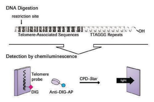
Telomeres are specific structures found at the end of chromosomes in eukaryotes. In human chromosomes, the telomeres consist of thousands of copies of 6 base repeats (TTAGGG)(1-3). It has been suggested that telomeres protect chromosome ends since damaged chromosomes lacking telomeres undergo fusion, rearrangement and translocation (2). In somatic cells, telomere length is progressively shortened with each cell division both in vivo and in vitro (4-7) due to the inability of the DNA polymerase complex to synthesize the very 5′ end of the lagging strand (8,9).
Telomerase is a ribonucleoprotein that synthesizes and directs the telomeric repeats onto the 3′ end of existing telomeres using its RNA component as a template (10-14). Telomerase activity has been shown to be specifically expressed in immortal cells, cancer and germ cells (15,16) where it compensates for telomere shortening during DNA replication and thus stabilizes telomere length (7,17). These observations have led to a hypothesis that telomere length may function as a "mitotic clock" to sense the number of cell divisions and eventually signal replicative senescence or programmed cell death when a critical telomere length is achieved. Therefore, expression of telomerase activity in cancer cells may be a necessary and essential step for tumor development and progression (16,18-20). The causal relationship between expression of telomerase and telomere length stabilization and the extension of the life span of the human cell has recently been reported (21).
The development of a sensitive and efficient PCR-based telomerase activity detection method, TRAP (Telomeric Repeat Amplification Protocol) (15, 22), has made possible large scale surveys of telomerase activity in human cells and tissues (15, 23-29). To date, telomerase activity has been detected in over 85% of all tumors tested spanning more than 20 different types of cancers (30-31).
The TRAPeze™ RT Telomerase Detection Kit is a highly sensitive in vitro assay for the fluorometric detection and real time quantification of telomerase activity. It incorporates refinements to the original TRAP assay that were first introduced in the gel-based TRAPeze™ Telomerase Detection Kit (Cat. No. S7700) and adds the ability to quantitate telomerase activity using fluorescence energy transfer (ET) primers similar to the TRAPeze™ XL Telomerase Detection Kit (Cat. No. S7707). As in the original TRAPeze™ Kit, primer sequence modifications that reduce amplification artifacts and PCR controls for standard curve generation are included. In addition, both the TRAPeze™ RT and XL Kits use fluorescence energy transfer (ET) primers to generate fluorescently labeled TRAP products which permit nonisotopic, quantitative analysis of telomerase activity.
The unique design of these ET primers (Amplifluor® primers) allows detection and quantification of telomerase activity by directly measuring real time fluorescence emission in the reaction vessels. Since Amplifluor® primers will fluoresce only upon incorporation into the TRAP products, post-PCR sample manipulations such as electrophoretic gel or ELISA analyses are eliminated, thereby reducing the the risk of carry-over contamination. Quantitative analysis is not compromised when detection is performed in a high-throughput 96-well format unlike platforms utilizing a qualitative ELISA. Additionaly, an additional stand alone control is provided separately to assess PCR inhibitors that may be present in experimental samples. (Please see product insert for references).
The TRAPeze™ RT Telomerase Detection Kit is a highly sensitive in vitro assay for the fluorometric detection and real time quantification of telomerase activity. It incorporates refinements to the original TRAPeze™ assay and adds the ability to quantitate telomerase activity using fluorescence energy transfer (ET) primers.
포장
The kit provides enough reagents to perform 224 TRAPeze™ RT reactions.
성분
CHAPS Lysis Buffer (13.5mL)
5X TRAPeze™ RT Reaction Mix (1.12mL)
5X TRAPeze™ Control Reaction Mix (1.12mL)
PCR - Grade Water (8.2mL)
TSR8* (quantitation control template) (45 μL)
TSK* (pcr inhibition/normalization control) (45 μL)
Control Cell Pellet (Telomerase positive cells; 106 cells)
* Caution - refer to Sec. II. Kit Components, Precautions in product insert.
저장 및 안정성
1. CHAPS Lysis Buffer - 15°C to -25°C
2. 5X TRAPeze™ RT Reaction Mix -15°C to -25°C
3. 5X TRAPeze™ Control Reaction Mix 2°C to 8°C
4. PCR - Grade Water - 15°C to -25°C
5. TSR8 -15°C to -25°C
6. TSK -15°C to -25°C
7. Control Cell Pellet -75°C to -85°C
2. 5X TRAPeze™ RT Reaction Mix -15°C to -25°C
3. 5X TRAPeze™ Control Reaction Mix 2°C to 8°C
4. PCR - Grade Water - 15°C to -25°C
5. TSR8 -15°C to -25°C
6. TSK -15°C to -25°C
7. Control Cell Pellet -75°C to -85°C
법적 정보
ABI PRISM is a registered trademark of Applera Corporation or its subsidiaries in the US and/or certain other countries
Amplifluor is a registered trademark of Merck KGaA, Darmstadt, Germany
CHEMICON is a registered trademark of Merck KGaA, Darmstadt, Germany
Opticon is a trademark of Bio-Rad Laboratories, Inc.
TRAPEZE is a trademark of Merck KGaA, Darmstadt, Germany
iCycler is a registered trademark of Bio-Rad
신호어
Warning
유해 및 위험 성명서
예방조치 성명서
Hazard Classifications
Aquatic Chronic 3 - Met. Corr. 1
Storage Class Code
8B - Non-combustible corrosive hazardous materials
Flash Point (°F)
Not applicable
Flash Point (°C)
Not applicable
시험 성적서(COA)
제품의 로트/배치 번호를 입력하여 시험 성적서(COA)을 검색하십시오. 로트 및 배치 번호는 제품 라벨에 있는 ‘로트’ 또는 ‘배치’라는 용어 뒤에서 찾을 수 있습니다.
Cullin 5 regulates cortical layering by modulating the speed and duration of Dab1-dependent neuronal migration.
Sim?? S, Jossin Y, Cooper JA
The Journal of Neuroscience null
Sandra Donnini et al.
FASEB journal : official publication of the Federation of American Societies for Experimental Biology, 24(7), 2385-2395 (2010-03-09)
Cerebral amyloid angiopathy (CAA) caused by amyloid beta (Abeta) deposition around brain microvessels results in vascular degenerative changes. Antiangiogenic Abeta properties are known to contribute to the compromised cerebrovascular architecture. Here we hypothesize that Abeta peptides impair angiogenesis by causing
A Static Magnetic Field Inhibits the Migration and Telomerase Function of Mouse Breast Cancer Cells.
Zhu Fan et al.
BioMed research international, 2020, 7472618-7472618 (2020-05-29)
Static magnetic field (SMF) has a potential as a cancer therapeutic modality due to its specific inhibitory effects on the proliferation of multiple cancer cells. However, the underlying mechanism remains unclear, and just a few studies have examined the effects
Kim E Innes et al.
Journal of Alzheimer's disease : JAD, 66(3), 947-970 (2018-10-16)
Telomere length (TL), telomerase activity (TA), and plasma amyloid-β (Aβ) levels have emerged as possible predictors of cognitive decline and dementia. To assess the: 1) effects of two 12-week relaxation programs on TL, TA, and Aβ levels in adults with
Sadia Mohsin et al.
Circulation research, 113(10), 1169-1179 (2013-09-21)
Myocardial function is enhanced by adoptive transfer of human cardiac progenitor cells (hCPCs) into a pathologically challenged heart. However, advanced age, comorbidities, and myocardial injury in patients with heart failure constrain the proliferation, survival, and regenerative capacity of hCPCs. Rejuvenation
자사의 과학자팀은 생명 과학, 재료 과학, 화학 합성, 크로마토그래피, 분석 및 기타 많은 영역을 포함한 모든 과학 분야에 경험이 있습니다..
고객지원팀으로 연락바랍니다.