
추천 제품


Quality Level
사용
sufficient for 1000 tests
sufficient for 200 tests
포장
pkg of 1 96-well plate(s)
제조업체/상표
Calbiochem®
저장 조건
OK to freeze
avoid repeated freeze/thaw cycles
입력
sample type intact cells
sample type intact cells
검출 방법
colorimetric
배송 상태
wet ice
저장 온도
−20°C
일반 설명
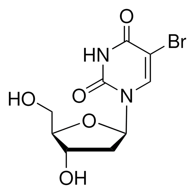
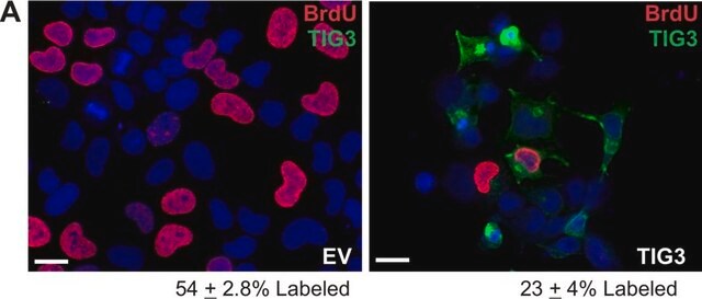
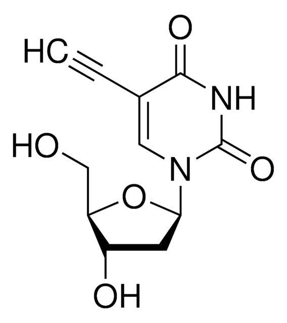
A non-isotopic enzyme immunoassay for the quantification of cell proliferation.Evaluation of cell cycle progression is essential for investigations in many scientific fields. Measurement of [3H] thymidine incorporation as cells enter S phase has long been the traditional method for the detection of cell proliferation. Subsequent quantification of [3H] thymidine is performed by scintillation counting or autoradiography. This technology is slow, labor intensive and has several limitations including the handling and disposal of radioisotopes and the necessity of expensive equipment. A well-established alternative to [3H] thymidine uptake has been demonstrated by numerous investigators. In these methods bromodeoxyuridine (BrdU), a thymidine analog replaces [3H] thymidine. BrdU is incorporated, into newly synthesized DNA strands of actively proliferating cells. Following partial denaturation of double stranded DNA, BrdU is detected immunochemically allowing the assessment of the population of cells, which are actively synthesizing DNA. The Calbiochem® BrdU Cell Proliferation Assay involves incorporation of BrdU into cells cultured in plates and BrdU immunolabeling using the cell layer as the solid phase. The resultant assay is sensitive, rapid, easy to perform and applicable to high sample throughput. In addition to evaluation of cell proliferation, information such as cell number, morphology and analysis of cellular antigens can be obtained from a single culture.
BrdU Cell Proliferation Assay is a non-isotopic immunoassay for quantification of BrdU incorporation into newly synthesized DNA of actively proliferating cells. It is sensitive, rapid, and easy to perform.
Note: 1 T = 1 test.
This proliferation assay is a non-isotopic immunoassay for quantification of BrdU incorporation into newly synthesized DNA of actively proliferating cells.
성분
BrdU Label, Fixative/Denaturing Solution, BrdU Antibody, Antibody Diluent, Peroxidase Goat Anti-Mouse IgG, Conjugate Diluent, Substrate, Plate Wash Concentrate, Stop Solution, and a user protocol.
경고
Toxicity: Multiple Toxicity Values, refer to MSDS (O)
규격
Assay Time: 3 h
원리
The Calbiochem® BrdU Cell Proliferation Assay is a non-isotopic immunoassay for the quantitation of bromodeoxyuridine incorporation into newly synthesized DNA of actively proliferating cells.
저장 및 안정성
Upon arrival store the entire contents of the kit at -20°C, in a non-frostfree freezer.
Prior to use:
1. Remove the Fixative/Denaturing Solution and place at room temperature for at least 4 h prior to use. Precipitates that may occur while cold should go back into solution.
2. Reconstitute the Peroxidase Goat Anti-Mouse IgG with the appropriate volume of 1X PBS.
• For the 200 test kit, reconstitute Peroxidase Goat Anti-Mouse IgG in 125 µl of 1X PBS. Use Conjugate Diluent for any further lot-specific dilution.
• For the 1000 test kit, reconstitute Peroxidase Goat Anti-Mouse IgG in 250 µl of 1X PBS. Use Conjugate Diluent for any further lot-specific dilution.
Allow solution to stand at room temperature for 10 min. (or until solution is clear). Divide into small aliquots; aliquots not to be used that same day should be stored at -20°C in a non-frostfree freezer.
Once the kit components have been thawed for use:
• The BrdU label and 100X Anti-BrdU Antibody should be divided into small aliquots and stored at -20°C in a non-frostfree freezer with the Peroxidase Goat Anti-Mouse IgG; avoid multiple freeze/thaw cycles.
• All the other kit components (Substrate, Antibody Diluent, Conjugate Diluent, 20X Plate Wash Concentrate, Fixative/Denaturing Solution, Stop Solution) should be stored at 4°C.
기타 정보
Due to the nature of the Hazardous Materials in this shipment, additional shipping charges may be applied to your order. Certain sizes may be exempt from the additional hazardous materials shipping charges. Please contact your local sales office for more information regarding these charges.
Hardonk, M.J. and Harms, G. 1990. Acta histochemica, Suppl.39, 99.
Muir, D., et al. 1990. Analytical Biochemistry185, 377.
Lanier, T. L., et al. 1989. Carcinogenesis10, 1341.
Magaud, J.-P., et al. 1988. J. Immunol. Methods106, 95.
Collins, S.J. 1987. Blood70, 1233.
Porstmann, T., et al. 1985. J. Immunol. Methods82, 169.
Raza, A., et al. 1985. Cytometry6, 633.
Morstyn, G., et al. 1983. J. Clin. Invest.72, 1844.
Gratzner, H.G. 1982. Science218, 474.
법적 정보
CALBIOCHEM is a registered trademark of Merck KGaA, Darmstadt, Germany


신호어
Danger
유해 및 위험 성명서
Hazard Classifications
Carc. 2 - Eye Irrit. 2 - Flam. Liq. 2 - Met. Corr. 1 - Muta. 1B - Repr. 2 - Skin Irrit. 2 - Skin Sens. 1
Storage Class Code
3 - Flammable liquids
Flash Point (°F)
69.8 °F
Flash Point (°C)
21 °C
시험 성적서(COA)
제품의 로트/배치 번호를 입력하여 시험 성적서(COA)을 검색하십시오. 로트 및 배치 번호는 제품 라벨에 있는 ‘로트’ 또는 ‘배치’라는 용어 뒤에서 찾을 수 있습니다.
이미 열람한 고객

Md Shahaduzzaman et al.
Age (Dordrecht, Netherlands), 35(6), 2071-2087 (2012-12-25)
Neurogenesis occurs throughout life but significantly decreases with age. Human umbilical cord blood mononuclear cells (HUCB MNCs) have been shown to increase the proliferation of neural stem cells (NSCs) in the dentate gyrus (DG) of the hippocampus and the subgranular
Mathiyazhagan Rengasamy et al.
The Indian journal of medical research, 144(6), 852-864 (2017-05-06)
Administration of ex vivo-expanded human bone marrow-derived mesenchymal stromal cells (hBMMSC) obtained from single donors has shown therapeutic benefits in both preclinical and clinical studies. In this study, the safety, toxicity and biodistribution profiles of a pooled hBMMSC population, produced
Igor Martianov et al.
PloS one, 9(2), e87365-e87365 (2014-02-06)
Collagen 6A3 (Col6a3), a component of extracellular matrix, is often up-regulated in tumours and is believed to play a pro-oncogenic role. However the mechanisms of its tumorigenic activity are poorly understood. We show here that Col6a3 is highly expressed in
Tamasa De et al.
Journal of clinical medicine, 8(9) (2019-09-19)
The majority of the cancer-associated deaths is due to metastasis-the spread of tumors to other organs. Circulating tumor cells (CTCs), which are shed from the primary tumor into the circulation, serve as precursors of metastasis. CTCs have now gained much
Hui Gao et al.
Scientific reports, 9(1), 9949-9949 (2019-07-11)
Nicotinamide phosphoribosyltransferase (NAMPT) upregulation in human pulmonary artery endothelial cells (hPAECs) is associated with pulmonary arterial hypertension (PAH) progression and pulmonary vascular remodeling. The underlying mechanisms regulating NAMPT expression are still not clear. In this study, we aimed to study
자사의 과학자팀은 생명 과학, 재료 과학, 화학 합성, 크로마토그래피, 분석 및 기타 많은 영역을 포함한 모든 과학 분야에 경험이 있습니다..
고객지원팀으로 연락바랍니다.