추천 제품
일반 설명
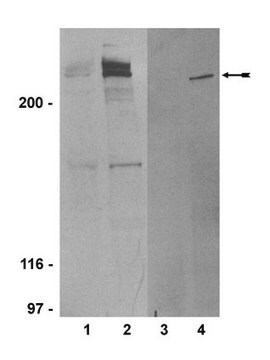
Anti-ATM (Ab-3) (819-844), rabbit polyclonal, recognizes the ~350 kDa ATM protein in Daudi and HeLa cells. It is validated for Western blotting and immunoprecipitation.
Purified rabbit polyclonal antibody. Recognizes the ~350 kDa ATM protein.
Recognizes the ~350 kDa ATM protein in Daudi and HeLa cells.
면역원
Human
a synthetic peptide (CKSLASFIKKPFDRGEVESMEDDTNG) corresponding to amino acids 819-844 of human ATM
애플리케이션
Immunoblotting (2 µg/ml, see comments)
Immunoprecipitation (2 µg)
Immunoprecipitation (2 µg)
포장
Please refer to vial label for lot-specific concentration.
경고
Toxicity: Standard Handling (A)
물리적 형태
In 50 mM sodium phosphate buffer, 0.2% gelatin.
분석 메모
Negative Control
GM02052A cells
GM02052A cells
Positive Control
Daudi or HeLa cells
Daudi or HeLa cells
기타 정보
For immunoblotting use 100-150 µg cell lysate on a 5% acrylamide gel (SDS/PAGE) and transferred to nitrocellulose using semi-dry transfer at 9V constant voltage for 2 h. Detection of antibody/antigen complexes is done using HRP conjugated goat anti-rabbit IgG at 25 ng/ml (Cat. No. DC03L).
1. Anti-ATM (Ab-3) Rabbit pAb immunoblots and immunoprecipitates a 350 kDa protein in lysates of normal and transformed cells or tumor lines derived from individuals homozygous wild type for the ATM gene; whereas in cells derived from patients with Ataxia-Telangiectasia and carrying homozygous inactivating mutations in ATM, no 350 kDa protein is detected.
2. Anti-ATM (Ab-3) Rabbit pAb may non-specifically detect smaller molecular weight proteins present in both ATM mutant and wild type cells. Careful titering of primary and secondary antibodies is recommended.
3. Immunoblotting of p350ATM requires loading 100-150 µg cell lysate on low percentage acrylamide gels (5%) (SDS/PAGE) with electrophoresis performed until the 200 kDa molecular weight marker has migrated halfway through the gel. Semi-dry electrophoretic transfer is for 2 h at 9V constant voltage. Tank transfer is overnight at 40 V constant voltage.
4. To transfer the gel for blotting, lay a dry piece of Whatman 3MM Chromatography paper over the wet gel. Carefully peel the 3MM paper and gel off the glass plate and immerse, gel side up, in transfer buffer until 3MM paper is thoroughly wet. Remove bubbles by rolling a pipette across the surface of the gel.
5. To confirm detection of p350ATM, a cell line carrying a truncating mutation in ATM should be used as a negative control. Cell lines from A-T patients which show no detectable band at 350 kDa, can be obtained from the National Institute of General Medical Sciences Human Genetic Mutant Cell Repository at the Coriell Institute for Medical Research, 401 Haddon Avenue, Camden, NJ 08103.
6. For immunoprecipitations, prepare nuclear lysates as described. Immunoprecipitate p350ATM using 2 µg of Anti-ATM (Ab-3) Rabbit pAb and Protein A-Agarose (Cat. No. IP06). Detection can be after metabolic labeling with 35S methionine followed by autoradiography, or alternatively, immunoprecipitated proteins can be displayed on 5% SDS/PAGE, transferred to nitrocellulose and then blotted as above using Anti-ATM (Ab-3).
Friedberg, E.C., et al. 1995. Amer. Soc. of Microbiolgy (meeting report), Wash. D.C.
Meyn, S.M. 1995. Cancer Res.55, 5991.
Paules, R.S., et al. 1995. Cancer Res.55, 1763.
Savitsky, K., et al. 1995. Science268, 1749.
Savitsky, K., et al. 1995. Hum. Molec. Genet.4, 2025.
Zakian, V., 1995. Cell82, 685.
Beamish, H., et al. 1993. Rad. Res.138, 130.
Kastan, M.B., et al. 1992. Cell71, 587.
Painter, R.B. and Young, B.R. 1980. Proc. Natl. Acad. Sci. USA77, 7315.
법적 정보
CALBIOCHEM is a registered trademark of Merck KGaA, Darmstadt, Germany
적합한 제품을 찾을 수 없으신가요?
당사의 제품 선택기 도구.을(를) 시도해 보세요.
Storage Class Code
10 - Combustible liquids
WGK
nwg
Flash Point (°F)
Not applicable
Flash Point (°C)
Not applicable
시험 성적서(COA)
제품의 로트/배치 번호를 입력하여 시험 성적서(COA)을 검색하십시오. 로트 및 배치 번호는 제품 라벨에 있는 ‘로트’ 또는 ‘배치’라는 용어 뒤에서 찾을 수 있습니다.
Yingli Sun et al.
Molecular and cellular biology, 27(24), 8502-8509 (2007-10-10)
The ATM protein kinase is essential for cells to repair and survive genotoxic events. The activation of ATM's kinase activity involves acetylation of ATM by the Tip60 histone acetyltransferase. In this study, systematic mutagenesis of lysine residues was used to
Norvin D Fernandes et al.
The Journal of biological chemistry, 282(22), 16577-16584 (2007-04-12)
The ATM protein kinase is mutated in ataxia telangiectasia, a genetic disease characterized by defective DNA repair, neurodegeneration, and growth factor signaling defects. The activity of ATM kinase is activated by DNA damage, and this activation is required for cells
Meilin Wang et al.
Nature communications, 5, 4901-4901 (2014-09-25)
ATM- and RAD3-related (ATR)/Chk1 and ataxia-telangiectasia mutated (ATM)/Chk2 signalling pathways play critical roles in the DNA damage response. Here we report that the E3 ubiquitin ligase Smurf1 determines cell apoptosis rates downstream of DNA damage-induced ATR/Chk1 signalling by promoting degradation
Yingli Sun et al.
Nature cell biology, 11(11), 1376-1382 (2009-09-29)
DNA double-strand break (DSB) repair involves complex interactions between chromatin and repair proteins, including Tip60, a tumour suppressor. Tip60 is an acetyltransferase that acetylates both histones and ATM (ataxia telangiectasia mutated) kinase. Inactivation of Tip60 leads to defective DNA repair
S Faderl et al.
Leukemia, 16(6), 1045-1052 (2002-06-01)
It has been suggested that the expansion of the leukemic cells in chronic lymphocytic leukemia (CLL) is due to dysregulation of pathways of programmed cell death (apoptosis) rather than cell proliferation, although differences may exist in early vs late and
자사의 과학자팀은 생명 과학, 재료 과학, 화학 합성, 크로마토그래피, 분석 및 기타 많은 영역을 포함한 모든 과학 분야에 경험이 있습니다..
고객지원팀으로 연락바랍니다.


