추천 제품
생물학적 소스
mouse
항체 형태
purified immunoglobulin
항체 생산 유형
primary antibodies
클론
4C11, monoclonal
종 반응성
human, monkey, Syrian hamster, rat, mouse
포장
antibody small pack of 25 μg
기술
ChIP: suitable
immunofluorescence: suitable
immunoprecipitation (IP): suitable
western blot: suitable
동형
IgG2aκ
NCBI 수납 번호
UniProt 수납 번호
타겟 번역 후 변형
unmodified
유전자 정보
human ... LMNA(4000)
일반 설명
Prelamin-A/C (UniProt: P02545) is encoded by the LMNA (also known as LMN1) gene (Gene ID: 4000) in human. It is cleaved into Lamin-A/C (also known as 70 kDa Lamin, Renal carcinoma antigen NY-REN-32). Lamins are components of the nuclear lamina that provide a framework for the nuclear envelope and may also interact with chromatin. Lamin A and C are present in equal amounts in the lamina of mammals. Plays an important role in nuclear assembly, chromatin organization, nuclear membrane and telomere dynamics. Lamin A is initially synthesized as prelamin A that undergoes several modifications in the carboxyl terminal region that allow incorporation of prelamin A into the nuclear envelope and its subsequent processing into the mature lamin A. Cleavage of 15 residues (aa 647-662) by ZMPSTE24/FACE1 generates the final protein product. Unlike mature lamin A, prelamin A accumulates as discrete and localized foci at the nuclear periphery. Prelamin-A/C can accelerate smooth muscle cell senescence. It can act to disrupt mitosis and induce DNA damage in vascular smooth muscle cells (VSMCs), leading to mitotic failure, genomic instability, and premature senescence. Mutations in LMNA gene are known to cause Emery-Dreifuss muscular dystrophy that is characterized by weakness and atrophy of muscle without involvement of the nervous system. Some mutations have also been linked to familial type of lipodystrophy characterized by the loss of subcutaneous adipose tissue in the lower parts of the body. (Ref.: Casasola, A., et al. (2016). Nucleus 7(1); 84-102).
특이성
Clone 4C11 detects Lamin A/C at the nuclear lamina in multiple species.
면역원
Purified protein corresponding to the Ig-fold of human Lamin A/C.
애플리케이션
Anti-Lamin A/C, clone 4C11, Cat. No. MABT1341, is a highly specific mouse monoclonal antibody that targets Lamin A/C and has been tested for use in Immunofluorescence, Immunoprecipitation, Chromatin Immunoprecipitation (ChIP), and Western Blotting.
Research Category
Cell Structure
Cell Structure
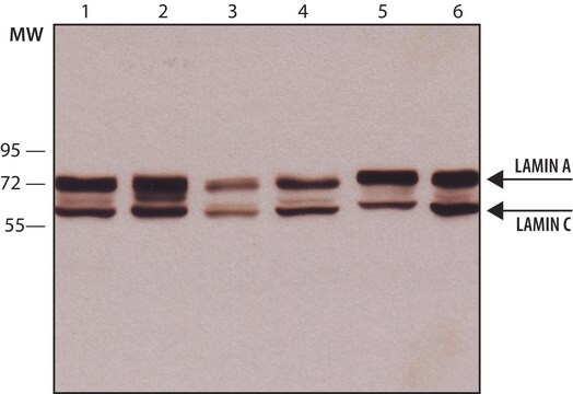
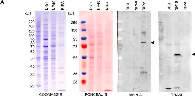
Western Blotting Analysis: A representative lot detected Lamin A/C in Western Blotting applications (Gesson, K., et. al. (2016). Genome Res. 26(4):462-73; Roblek, M., et. al. (2010). PLoS One. 5(5):e10604).
Dot Blot Analysis: A representative lot detected Lamin A/C in Dot Blot applications (Roblek, M., et. al. (2010). PLoS One. 5(5):e10604).
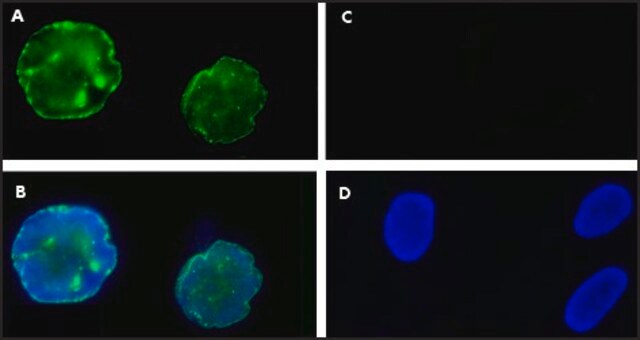
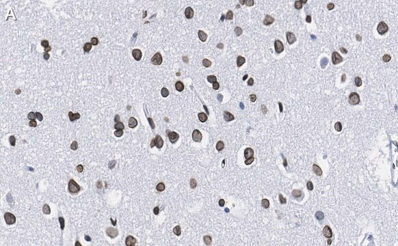
Immunocytochemistry Analysis: A 1:1,000 dilution from a representative lot detected Lamin A/C in HeLa cells.
Western Blotting Analysis: A representative lot detected Lamin A/C in WB used to demonstrate high titration and excellent properties of clone 4C11 (Courtesy of Marie lang, M.D., Stefan Schuchner, Ph.D. and Egon Ogris, M.D., Medical University of Vienna, Austria).
Immunofluorescence Analysis: A representative lot detected the Lamin A/C species at the nuclear lamina of HeLa cells (Courtesy of Marie Lang, M.D., Stefan Schuchner, Ph.D. and Egon Ogris, M.D., Medical University of Vienna, Austria).
Immunoprecipitation Analysis: A representative lot detected Lamin A/C in WB used to demonstrate high titration and excellent properties of clone 4C11 (Courtesy of Marie Lang, M.D., Stefan Schuchner, Ph.D. and Egon Ogris, M.D., Medical University of Vienna, Austria).
Chromatin Immunoprecipitation (ChIP) Analysis: A representative lot detected Lamin A/C in Chromatin Immunoprecipitation applications (Gesson, K., et. al. (2016). Genome Res. 26(4):462-73).
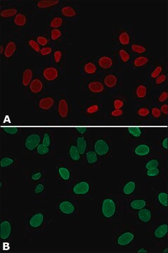
Immunofluorescence Analysis: A representative lot detected Lamin A/C in Immunofluorescence applications (Roblek, M., et. al. (2010). PLoS One. 5(5):e10604).
Immunoprecipitation Analysis: A representative lot immunoprecipitated Lamin A/C in Immunoprecipitation applications (Gesson, K., et. al. (2016). Genome Res. 26(4):462-73).
품질
Evaluated by Western Blotting in HeLa cell lysate.
Western Blotting Analysis: 0.2 µg/mL of this antibody detected Lamin A/C in HeLa cell lysate.
Western Blotting Analysis: 0.2 µg/mL of this antibody detected Lamin A/C in HeLa cell lysate.
표적 설명
74 and 65 kDa observed. 74.14 kDa and 65.14 kDa calculated for Lamin A and C, respectively. Uncharacterized bands may be observed in some lysate(s).
물리적 형태
Format: Purified
Protein G purified
Purified mouse monoclonal antibody IgG2a in buffer containing 0.1 M Tris-Glycine (pH 7.4), 150 mM NaCl with 0.05% sodium azide.
저장 및 안정성
Stable for 1 year at 2-8°C from date of receipt.
기타 정보
Concentration: Please refer to lot specific datasheet.
면책조항
Unless otherwise stated in our catalog or other company documentation accompanying the product(s), our products are intended for research use only and are not to be used for any other purpose, which includes but is not limited to, unauthorized commercial uses, in vitro diagnostic uses, ex vivo or in vivo therapeutic uses or any type of consumption or application to humans or animals.
적합한 제품을 찾을 수 없으신가요?
당사의 제품 선택기 도구.을(를) 시도해 보세요.
Storage Class Code
12 - Non Combustible Liquids
WGK
WGK 1
Flash Point (°F)
Not applicable
Flash Point (°C)
Not applicable
시험 성적서(COA)
제품의 로트/배치 번호를 입력하여 시험 성적서(COA)을 검색하십시오. 로트 및 배치 번호는 제품 라벨에 있는 ‘로트’ 또는 ‘배치’라는 용어 뒤에서 찾을 수 있습니다.
자사의 과학자팀은 생명 과학, 재료 과학, 화학 합성, 크로마토그래피, 분석 및 기타 많은 영역을 포함한 모든 과학 분야에 경험이 있습니다..
고객지원팀으로 연락바랍니다.