추천 제품
생물학적 소스
mouse
항체 형태
purified antibody
항체 생산 유형
primary antibodies
클론
2G10.2, monoclonal
분자량
~30 kDa (Uncharacterized bands may be observed in some lysates.)
정제법
using protein G
종 반응성
human, mouse, rat
기술
immunofluorescence: suitable
western blot: suitable
동형
IgG2bκ
NCBI 수납 번호
UniProt 수납 번호
유전자 정보
mouse ... TPM3(117557)
일반 설명
Tropomyosin alpha-3 chain (UniProt: Q63610; also known as Gamma-tropomyosin, Tropomyosin-3, Tropomyosin-5) is encoded by the Tpm3 (also known as Tpm-5, Tpm5) gene (Gene ID: 117557) in rat. The TPM3 gene codes for the slow-twitch skeletal muscle isoform ( s Tm) and at least 9 LMW cytoskeletal isoforms referred to as Tm5NM1 to Tm5NM11. Tropomyosins are dimers of coiled-coil proteins that provide stability to actin filaments and regulate access of other actin-binding proteins. In muscle cells, they regulate muscle contraction by controlling the binding of myosin heads to the actin filament. In non-muscle cells tropomyosins are implicated in stabilizing cytoskeleton actin filaments. Tropomyosins have been implicated in the pathogenesis of cancer where high molecular weight isoforms are consistently down-regulated in transformed cells, while malignant cells display an increased reliance on low molecular weight isoforms. Tropomyosin 3 is a homodimeric protein that can form heterodimers with a beta (TPM2) chain. Its coiled coil structure is formed by 2 polypeptide chains. (Ref.: Glass, JJ et al. (2015). BMC Cancer. 15; 712).
특이성
Clone 2G10.2 is a mouse monoclonal antibody that specifically detects Tpm3.1 and Tpm3.2. It targets an epitope within 27 amino acids from the C-terminal region.
면역원
Diphtheria Toxoid Carrier Protein-conjugated linear peptide corresponding to 27 amino acids from the C-terminal region of rat Tpm3.1 and Tpm3.2
애플리케이션
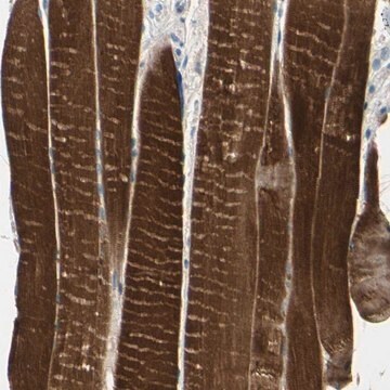
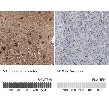
Western Blotting Analysis: 1 µg/mL from a representative lot detected Tropomyosin 3 in human lung tissue lysate.
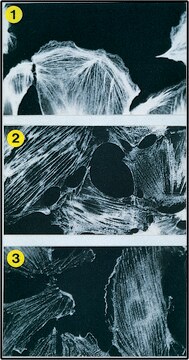
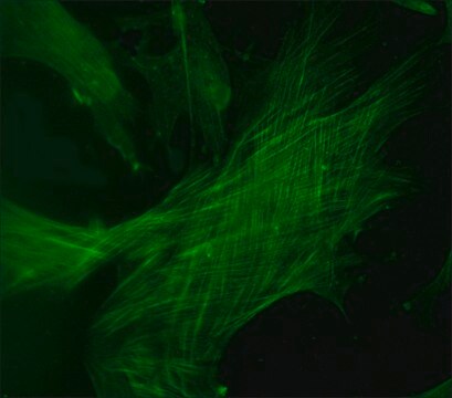
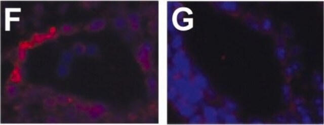
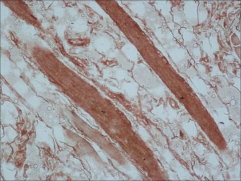
Immunofluorescence Analysis: A representative lot detected Tropomyosin 3 in Immunofluorescence applications (Schevzov, G., et. al. (2011). Bioarchitecture. 1(4):135-164).
Western Blotting Analysis: A representative lot detected Tropomyosin 3 in Western Blotting applications (Schevzov, G., et. al. (2011). Bioarchitecture. 1(4):135-164).
품질
Evaluated by Western Blotting in mouse liver tissue lysate. Western Blotting Analysis: 1 µg/mL of this antibody detected Tropomyosin 3 in mouse liver tissue lysate.
물리적 형태
Purified mouse monoclonal antibody IgG2b in buffer containing 0.1 M Tris-Glycine (pH 7.4), 150 mM NaCl with 0.05% sodium azide.
저장 및 안정성
Stable for 1 year at 2-8°C from date of receipt.
기타 정보
Concentration: Please refer to lot specific datasheet.
면책조항
Unless otherwise stated in our catalog or other company documentation accompanying the product(s), our products are intended for research use only and are not to be used for any other purpose, which includes but is not limited to, unauthorized commercial uses, in vitro diagnostic uses, ex vivo or in vivo therapeutic uses or any type of consumption or application to humans or animals.
적합한 제품을 찾을 수 없으신가요?
당사의 제품 선택기 도구.을(를) 시도해 보세요.
시험 성적서(COA)
제품의 로트/배치 번호를 입력하여 시험 성적서(COA)을 검색하십시오. 로트 및 배치 번호는 제품 라벨에 있는 ‘로트’ 또는 ‘배치’라는 용어 뒤에서 찾을 수 있습니다.
자사의 과학자팀은 생명 과학, 재료 과학, 화학 합성, 크로마토그래피, 분석 및 기타 많은 영역을 포함한 모든 과학 분야에 경험이 있습니다..
고객지원팀으로 연락바랍니다.