
추천 제품

생물학적 소스
mouse
Quality Level
항체 형태
purified immunoglobulin
항체 생산 유형
primary antibodies
클론
K65/35, monoclonal
종 반응성
rat
종 반응성(상동성에 의해 예측)
mouse (based on 100% sequence homology)
기술
immunohistochemistry: suitable
western blot: suitable
동형
IgG1κ
NCBI 수납 번호
UniProt 수납 번호
배송 상태
wet ice
타겟 번역 후 변형
unmodified
유전자 정보
rat ... Cntnap1(84008)
일반 설명
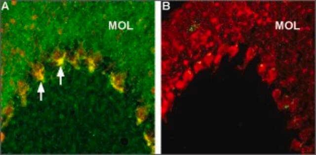
Contactin-associated protein 1(Caspr) is a single-pass type I membrane protein belonging to the neurexin family and is comprised of one F5/8 type C domain, one fibrinogen C-terminal domain, two EGF-like domains, and four laminin G-like domains. Caspr is well documented for its interaction with contactin, an interaction that is crucial to the transport of Caspr to the plasma membrane, the mylination of glial cells, and the generation of axoglial junctions. Studies also suggest that Caspr may play a regulatory role in the cell surface expression of contactin and its targeting to different axonal domains. Expression of Caspr is largely observed in the brain, mainly in CNS myelinated nerve fibers within paranodal axoglial junctions.
면역원
Recombinant protein corresponding to rat Caspr.
애플리케이션
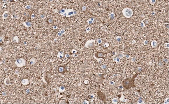
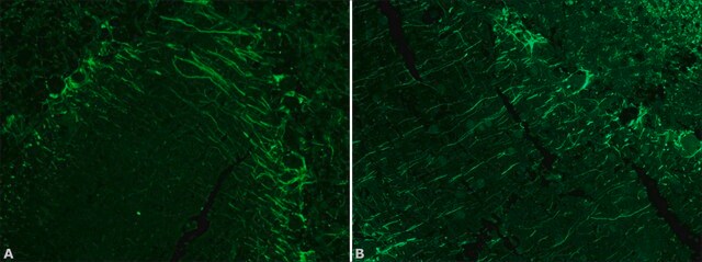
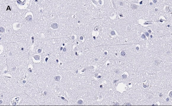
Anti-Caspr Antibody, clone K65/35 is an antibody against Caspr for use in IH & WB.
Immunohistochemistry Analysis: 1:300 dilution from a previous lot detected Caspr in rat cerebellum and rat hippocampus tissues.
Research Category
Neuroscience
Neuroscience
Research Sub Category
Signaling Neuroscience
Signaling Neuroscience
품질
Evaluated by Western Blot in rat brain membrane tissue lysate.
Western Blot Analysis: 0.5 µg/mL of this antibody detected Caspr on 10 µg of rat brain membrane tissue lysate.
Western Blot Analysis: 0.5 µg/mL of this antibody detected Caspr on 10 µg of rat brain membrane tissue lysate.
표적 설명
~ 220 kDa observed
물리적 형태
Format: Purified
Protein G
Purified mouse monoclonal IgG1κ in buffer containing 0.1 M Tris-Glycine (pH 7.4), 150 mM NaCl with 0.05% sodium azide.
저장 및 안정성
Stable for 1 year at 2-8°C from date of receipt.
분석 메모
Control
Rat brain membrane tissue lysate
Rat brain membrane tissue lysate
기타 정보
Concentration: Please refer to the Certificate of Analysis for the lot-specific concentration.
면책조항
Unless otherwise stated in our catalog or other company documentation accompanying the product(s), our products are intended for research use only and are not to be used for any other purpose, which includes but is not limited to, unauthorized commercial uses, in vitro diagnostic uses, ex vivo or in vivo therapeutic uses or any type of consumption or application to humans or animals.
적합한 제품을 찾을 수 없으신가요?
당사의 제품 선택기 도구.을(를) 시도해 보세요.
Storage Class Code
12 - Non Combustible Liquids
WGK
WGK 1
Flash Point (°F)
Not applicable
Flash Point (°C)
Not applicable
시험 성적서(COA)
제품의 로트/배치 번호를 입력하여 시험 성적서(COA)을 검색하십시오. 로트 및 배치 번호는 제품 라벨에 있는 ‘로트’ 또는 ‘배치’라는 용어 뒤에서 찾을 수 있습니다.
Annika Balraj et al.
Developmental neurobiology, 82(4), 308-325 (2022-04-12)
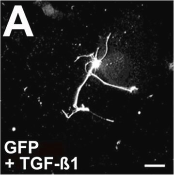
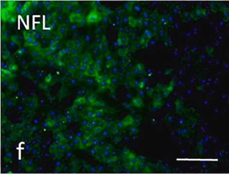
Retinal ganglion cells generate a pattern of action potentials to communicate visual information from the retina to cortical areas. Myelin, an insulating sheath, wraps axonal segments to facilitate signal propagation and when deficient, can impair visual function. Optic nerve development
Aníbal Sánchez-de la Torre et al.
Cell death & disease, 13(7), 585-585 (2022-07-08)
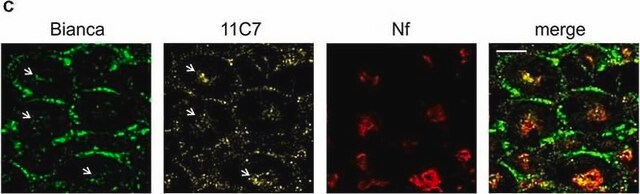
Cannabinoids are known to modulate oligodendrogenesis and developmental CNS myelination. However, the cell-autonomous action of these compounds on oligodendroglial cells in vivo, and the molecular mechanisms underlying these effects have not yet been studied. Here, by using oligodendroglial precursor cell
Evelien Fredrickx et al.
Glia, 68(6), 1148-1164 (2019-12-19)
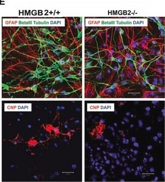
Myelin, one of the most important adaptations of vertebrates, is essential to ensure efficient propagation of the electric impulse in the nervous system and to maintain neuronal integrity. In the central nervous system (CNS), the development of oligodendrocytes and the
Di Chen et al.
Journal of neuroinflammation, 19(1), 112-112 (2022-05-17)
Microglia/macrophages are activated after cerebral ischemic stroke and can contribute to either brain injury or recovery by polarizing microglia/macrophage into distinctive functional phenotypes with pro- or anti-inflammatory properties. Interleukin-13 (IL-13) is an anti-inflammatory cytokine that regulates microglia/macrophage polarization toward an
자사의 과학자팀은 생명 과학, 재료 과학, 화학 합성, 크로마토그래피, 분석 및 기타 많은 영역을 포함한 모든 과학 분야에 경험이 있습니다..
고객지원팀으로 연락바랍니다.