
추천 제품


생물학적 소스
mouse
Quality Level
항체 형태
purified immunoglobulin
항체 생산 유형
primary antibodies
클론
6D1.5/A5, monoclonal
종 반응성
human
기술
ELISA: suitable
multiplexing: suitable
western blot: suitable
동형
IgG1
NCBI 수납 번호
UniProt 수납 번호
배송 상태
wet ice
타겟 번역 후 변형
unmodified
유전자 정보
human ... RNASE2(6036)
일반 설명
Non-secretory ribonuclease (EC 3.1.27.5; UniProt P10153; also known as Eosinophil-derived neurotoxin, Ribonuclease 2, Ribonuclease US, RNase 2, RNase UpI-2) is encoded by the RNASE2 (also known as EDN, RAF3, RNS2) gene (Gene ID 6036) in human. The major basic protein (MBP), eosinophil cationic protein (ECP), eosinophil derived neurotoxin (EDN) and eosinophil peroxidase (EPO), are eosinophil effector molecules implicated in host defense, immunoregulatory responses, and in allergic and inflammatory reactions. These highly cationic proteins are stored preformed within the eosinophil secondary granules and released in intact membrane-bound granules during cell lysis or through a complex system of vesiculotubular structures during “piecemeal necrosis”. While eosinophil count and eosinophil surface expression of the activation marker CD69 are significantly correlated with serum concentrations of MBP, EDN and EPO, studies show that they are not correlated with ECP level. On the other hand, eosinophilic disease patients with normal eosinophil count show significantly increased concentrations of MBP, ECP, EDN, and EPO compared to normal donors. The measurement of MBP, ECP, EDN, and EPO levels thus provides a better predictor than eosinophil count alone in disease diagnosis. EDN is produced with a signal peptide sequence (a.a. 1-27), the removal of which yields the mature (a.a. 28-161) protein.
특이성
Clone 6D1.5/A5 specifically reacted with EDN, but not other granule proteins by Western blotting analysis (Makiya, M.A., et al. (2014). J. Immunol. Methods. 411:11-22).
면역원
Purified human EDN.
애플리케이션
Anti-Eosinophil-Derived Neurotoxin Antibody, clone 6D1.5/A5 is an antibody against Eosinophil-Derived Neurotoxin for use in Western Blotting, ELISA, Multiplexing.
Research Category
Inflammation & Immunology
Inflammation & Immunology
Research Sub Category
Inflammation & Autoimmune Mechanisms
Inflammation & Autoimmune Mechanisms
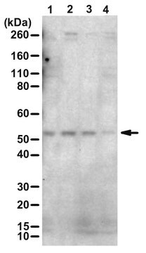
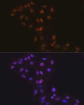
Western Blotting Analysis: 1.0 µg/mL from a representative lot detected eosinophil-derived neurotoxin in 50 µg of human liver and lung tissue lysates.
ELISA Analysis: A representative lot was employed as the capture antibody for the detection of purified human EDN, as well as EDN in serum samples from individuals with eosinophilic disorders by sandwich ELISA (Makiya, M.A., et al. (2014). J. Immunol. Methods. 411:11-22),
Multiplexing Analysis: A representative lot was employed as the capture antibody in a bead-based multiplex assay for the detection of purified human EDN as well as EDN in serum samples from healthy donors and individuals with eosinophilic disorders. Prior reduction and alkylation of the serum samples (by DTT and iodoacetamide, respectively) resulted in higher EDN levels (Makiya, M.A., et al. (2014). J. Immunol. Methods. 411:11-22),
품질
Evaluated by Western Blotting in human spleen tissue lysate.
Western Blotting Analysis: 1.0 µg/mL of this antibody detected eosinophil-derived neurotoxin in 50 µg of human spleen tissue lysate.
Western Blotting Analysis: 1.0 µg/mL of this antibody detected eosinophil-derived neurotoxin in 50 µg of human spleen tissue lysate.
표적 설명
~20 kDa observed. Target band appears larger than the calculated molecular weights of 18.35 kDa (pro-form) and 15.46 kDa (mature) due to glycosylation. Uncharacterized band(s) may appear in some lysates.
물리적 형태
Format: Purified
Protein G Purified
Purified mouse monoclonal IgG1 antibody in buffer containing 0.1 M Tris-Glycine (pH 7.4), 150 mM NaCl with 0.05% sodium azide.
저장 및 안정성
Stable for 1 year at 2-8°C from date of receipt.
기타 정보
Concentration: Please refer to lot specific datasheet.
면책조항
Unless otherwise stated in our catalog or other company documentation accompanying the product(s), our products are intended for research use only and are not to be used for any other purpose, which includes but is not limited to, unauthorized commercial uses, in vitro diagnostic uses, ex vivo or in vivo therapeutic uses or any type of consumption or application to humans or animals.
적합한 제품을 찾을 수 없으신가요?
당사의 제품 선택기 도구.을(를) 시도해 보세요.
WGK
nwg
Flash Point (°F)
does not flash
Flash Point (°C)
does not flash
시험 성적서(COA)
제품의 로트/배치 번호를 입력하여 시험 성적서(COA)을 검색하십시오. 로트 및 배치 번호는 제품 라벨에 있는 ‘로트’ 또는 ‘배치’라는 용어 뒤에서 찾을 수 있습니다.
자사의 과학자팀은 생명 과학, 재료 과학, 화학 합성, 크로마토그래피, 분석 및 기타 많은 영역을 포함한 모든 과학 분야에 경험이 있습니다..
고객지원팀으로 연락바랍니다.