추천 제품
생물학적 소스
mouse
항체 형태
purified antibody
항체 생산 유형
primary antibodies
클론
monoclonal
종 반응성
rat
제조업체/상표
Chemicon®
기술
immunohistochemistry: suitable
동형
IgM
배송 상태
dry ice
타겟 번역 후 변형
unmodified
특이성
Glutamate
The cross-reactivities were determined using an ELISA test by competition experiments with the following compounds:
Compound Cross-reactivity
Glutamate-G-BSA 1
Aspartate-G-BSA 1/100,000
GABA-G-BSA 1/100,000
Glutamate 1/100,000
Abbreviations:
(G) Glutaraldehyde
(BSA) Bovine Serum Albumin
NOTE: Antibody reactivity REQUIRES glutaraldehyde fixation thus some glutaraldehyde (0.5%-2.0%) needs to be included in the tissue fixation procedure inorder for the proper reactivity.
면역원
Glutamate-Glutaraldehyde-BSA.
애플리케이션
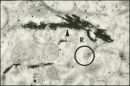
Anti-Glutamate Antibody detects level of Glutamate & has been published & validated for use in IH.
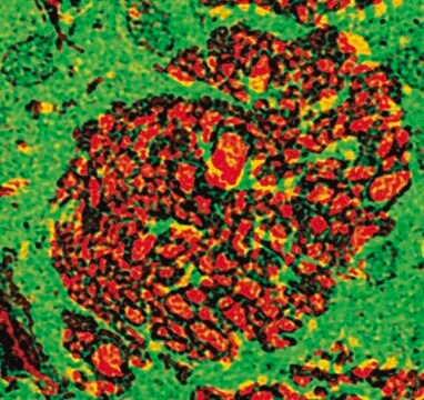
Immunohistochemistry: 1:2,500-1:10,000 using free floating sections by the PAP technique on rat hippocampus.
Optimal working dilutions must be determined by end user.
PROTOCOL for Glutamate Detection by Immunohisto/cytochemistry. Example for a rat brain.
1. SOLUTIONS TO BE PREPARED - Solution must be prepared as needed.
Solution A: 0.1M cacodylate, 10g/L sodium metabisulfite, pH 6.2.
Solution B: 0.1M cacodylate, 10g/L sodium metabisulfite, 3-5% glutaraldehyde, pH 7.5.
2. RAT PERFUSION - The rat is anaesthetized with sodium pentobarbital or Nembutal and perfused intracardially through the aorta using a pump with Solution A (30 mL): 150-300 mL/min, Solution B (500 mL): 150-300 mL/min.
3. POST FIXATION: 15 to 30 minutes in Solution B, then 4 soft washes in 0.05M Tris with 8.5 g/L sodium metabisulfite, pH 7.5 (Solution C) .
4. TISSUE SECTIONING: Vibratome or cryostat sections can be used.
5. REDUCTION STEP: Sections are reduced with Solution C containing 0.1M sodium borohydride for 10 minutes. The sections are washed 4 times in solution C without sodium borohydride.
6. APPLICATION OF GLUTAMATE ANTIBODY: Use a final dilution of 1:2,500-1:10,000 in Solution C containing 0.1% Triton X100 and 2% non-specific serum. Incubate 12 sections per 2 mL diluted antibody overnight, +2-8°C. Then wash the sections three times for 10 minutes each in Solution C. (Note that the antibody may be usable at a higher dilution. This should be explored to minimize the possibility of high background. Additionally, note that a change in the buffering system as indicated in the protocol may change the background and antibody recognition). The specific reaction is then revealed by PAP procedure.
7. SECOND ANTIBODY: Incubate the sections with a 1:50 to 1:200 dilution of goat anti-mouse in Solution B containing 1% non-specific serum for either 3 hrs at 20°C or 1-2 hr at 37°C. Then wash the sections, 3 times, for 10 minutes each with Solution C.
8. PAP: Incubate the sections with the appropriate dilution of peroxidase anti-peroxidase (for free floating method) in Solution C for 1-2 hours at 37°C. Then wash sections 3 times for 10 min each in solution C.
9. VISUALIZATION: The antigen-antibody complexes are visualized using DAB-4-HCl (25 mg/100 mL) (or other chromogen) in 0.05M Tris and filtrated; 0.05% hydrogen peroxide is added. Incubate the sections for 10 minutes at room temp. Stop the reaction by transferring the sections to 5 mL 0.05M Tris. Mount sections on chrome-alum coated slides. Dry overnight at 37°C. Rehydrate sections using conventional histological procedures. Coverslip using rapid mounting media.
For research use only; not for use as a diagnostic.
Research Category
Neuroscience
Neuroscience
Research Sub Category
Neurotransmitters & Receptors
Neuroinflammation & Pain
Neurotransmitters & Receptors
Neuroinflammation & Pain
물리적 형태
Format: Purified
Liquid in PBS containing 10mM sodium azide.
저장 및 안정성
Maintain at 2-8°C in undiluted aliquots for up to 6 months after date.
법적 정보
CHEMICON is a registered trademark of Merck KGaA, Darmstadt, Germany
면책조항
Unless otherwise stated in our catalog or other company documentation accompanying the product(s), our products are intended for research use only and are not to be used for any other purpose, which includes but is not limited to, unauthorized commercial uses, in vitro diagnostic uses, ex vivo or in vivo therapeutic uses or any type of consumption or application to humans or animals.
적합한 제품을 찾을 수 없으신가요?
당사의 제품 선택기 도구.을(를) 시도해 보세요.
Storage Class Code
12 - Non Combustible Liquids
WGK
WGK 2
Flash Point (°F)
Not applicable
Flash Point (°C)
Not applicable
시험 성적서(COA)
제품의 로트/배치 번호를 입력하여 시험 성적서(COA)을 검색하십시오. 로트 및 배치 번호는 제품 라벨에 있는 ‘로트’ 또는 ‘배치’라는 용어 뒤에서 찾을 수 있습니다.
Monoclonal antibody directed against glutaraldehyde conjugated glutamate and immunocytochemical applications in the rat brain.
Chagnaud, J L, et al.
Brain Research, 481, 175-180 (1989)
Spatial learning and memory deficit of low level polybrominated diphenyl ethers-47 in male adult rat is modulated by intracellular glutamate receptors.
Tang Yan,Li Xiang,Jiang Xuejun,Chen Chengzhi,Qi Youbin,Yu Xuelan,Liu Yang,Peng Changyan,Chen Hui
The Journal of Toxicological Sciences null
Anatomical characterization of a rabbit cerebellar eyeblink premotor pathway using pseudorabies and identification of a local modulatory network in anterior interpositus.
Gonzalez-Joekes, J; Schreurs, BG
The Journal of Neuroscience null
Lin Cheng et al.
Cell research, 24(6), 665-679 (2014-03-19)
Neural progenitor cells (NPCs) can be induced from somatic cells by defined factors. Here we report that NPCs can be generated from mouse embryonic fibroblasts by a chemical cocktail, namely VCR (V, VPA, an inhibitor of HDACs; C, CHIR99021, an
자사의 과학자팀은 생명 과학, 재료 과학, 화학 합성, 크로마토그래피, 분석 및 기타 많은 영역을 포함한 모든 과학 분야에 경험이 있습니다..
고객지원팀으로 연락바랍니다.