추천 제품
Quality Level
양식
liquid
IVD
for in vitro diagnostic use
기술
microbe id | staining: suitable
전이 온도
flash point 14 °C
density
0.83 g/cm3 at 20 °C
응용 분야
clinical testing
diagnostic assay manufacturing
hematology
histology
저장 온도
15-25°C
일반 설명
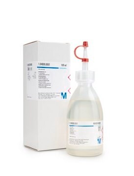
Papanicolaou′s solution 2a Orange G solution (OG6) - for cytology, is a ready-to-use solution used for human-medical cell diagnosis and serve the purpose of the cytological investigation of sample material of human origin, for example, cervical smears. Papanicolaou′s technique is the most used staining procedure for cytological specimens and is intended for the the staining of exfoliative cells in cytological specimens.
In the first step, the cell nuclei are stained either progressively or regressively with a hematoxylin solution, (Hematoxylin solution modified acc.to Gill II, (Product number 1.05175), Papanicolaou′s solution 1a Harris hematoxylin solution, (Product number 1.09253) or Papanicolaou′s solution 1b Hematoxylin solution S, (Product number 1.09254)). Nuclei are stained blue to dark violet. In the progressive hematoxylin staining method, staining is carried out to the endpoint, after which the slide is blued in tapwater. With the regressive method the material is over-stained and the excess of staining solution is removed by acid rinsing steps, followed by the bluing step. The structures of nuclei are more differentiated and better visible by the regressive method. The second staining step is cytoplasmic staining by orange staining solution, (Papanicolaou′s solution 2b Orange II solution, (Product number 1.106887) or Papanicolaou′s solution 2a Orange G solution (OG6), (Product number 1.06888)), especially for demonstration of mature and keratinized cells. The target structures are stained orange in different intensities. In the third staining step is used the so-called polychrome solution, a mixture of eosin, light green SF and Bismarck brown, Papanicolaou′s solution 3a polychromatic solution EA 31, (Product number 1.09271), Papanicolaou′s solution 3b polychromatic solution EA 50, (Product number 1.09272), Papanicolaou′s solution 3c polychromatic solution EA 65, (Product number 1.09270), or Papanicolaou′s solution 3d polychromatic solution EA 65, (Product number 1.09269). The polychrome solution is used for demonstration of differentiation of squamous cells. Papanicolaou′s solution 2a Orange G solution (OG6) gives a pale, yellow-orange staining result with mature and keratinized squamous cells.
The 500 ml bottle provides 1500 - 2000 stainings. This product is registered as IVD and CE marked. For more details, please see instructions for use (IFU). The IFU can be downloaded from this webpage.
분석 메모
Suitability for microscopy (Vaginal smear): passes test
Nuclei: blue to dark violet
Cyanophilic cytoplasm: blue-green
Eosinophilic cytoplasm: pink
Nuclei: blue to dark violet
Cyanophilic cytoplasm: blue-green
Eosinophilic cytoplasm: pink
신호어
Danger
유해 및 위험 성명서
Hazard Classifications
Eye Irrit. 2 - Flam. Liq. 2
Storage Class Code
3 - Flammable liquids
WGK
WGK 2
Flash Point (°F)
57.2 °F
Flash Point (°C)
14 °C
시험 성적서(COA)
제품의 로트/배치 번호를 입력하여 시험 성적서(COA)을 검색하십시오. 로트 및 배치 번호는 제품 라벨에 있는 ‘로트’ 또는 ‘배치’라는 용어 뒤에서 찾을 수 있습니다.
자사의 과학자팀은 생명 과학, 재료 과학, 화학 합성, 크로마토그래피, 분석 및 기타 많은 영역을 포함한 모든 과학 분야에 경험이 있습니다..
고객지원팀으로 연락바랍니다.


