詳細
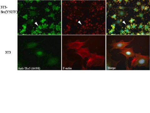
SH3 and PX domain-containing protein 2A (UniProt: Q5TCZ1; also known as Adapter protein TKS5, Five SH3 domain-containing protein, SH3 multiple domains protein 1, Tyrosine kinase substrate with five SH3 domains) is encoded by the SH3PXD2A (also known as FISH, KIAA0418, SH3MD1, TKS5) gene (Gene ID: 9644) in human. Adapter protein TKS5 is a scaffolding protein that is required for podosome formation, degradation of the extracellular matrix, and for invasion in some cancer cells. Three isoforms of TKS5 have been described that are produced by alternative splicing. TKS5 is a cytoplasmic protein in normal cells, but it localizes to podosomes in SRC-transformed cells. It is found in several cancer cell lines, particularly invasive breast carcinomas and melanomas. TKS5 contains one PX (phx homology) domain and five SH3 domains. The PX domain is required for podosome localization because of its ability to bind phosphoinosiltols, such as PtdIns3P and PtdIns(3,4)P2 and to lesser extent PtdIns5P and PtdIns(3,5)P2. Its binding with ADAM12, ADAM15 and ADAM19 is mediated by the fifth SH3 domain. The intramolecular interaction of the PX domain with the third SH3 domain maintains the protein in the cytoplasm and phosphorylation disrupts this interaction, resulting in the redistribution of the protein from cytoplasm to the perimembrane region.
特異性
Clone 13H6.3 detects SH3 and PX domain-containing protein 2A (adapter protein TKS5) in human cells. It targets and epitope with in 60 amino acids crom the C-terminal half.
免疫原
GST/His-tagged recombinant fragment corresponding to 60 amino acids from the C-terminal half of human SH3 and PX domain-containing protein 2A (Adapter protein TKS5).
アプリケーション
Research Category
細胞骨格
Anti-TKS5, clone 13H6.3, Cat. No. MABT336, ia a mouse monoclonal antibody that detects TKS5 and has been tested for use in Immunohistochemistry (Paraffin) and Western Blotting.
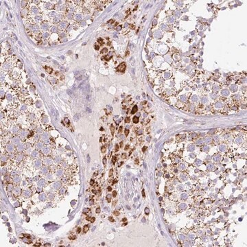
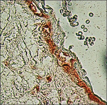
Immunohistochemistry Analysis: A 1:50 dilution from a representative lot detected TKS5 in human breast cancer tissue.
品質
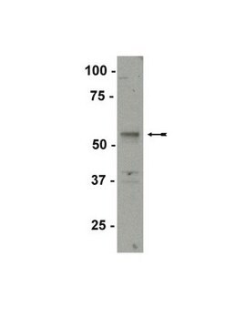
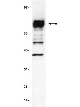
Evaluated by Western Blotting in SCC61 and SK-MEL28 cell lysates.
Western Blotting Analysis: 0.5 µg/mL of this antibody detected TKS5 in 10 µg of SCC61 and SK-MEL28 cell lysates.
ターゲットの説明
~160 kDa observed; 125.29 kda calculated. Uncharacterized bands may be observed in some lysate(s).
物理的形状
Protein G purified
Format: Purified
Purified mouse monoclonal antibody IgG1 in buffer containing 0.1 M Tris-Glycine (pH 7.4), 150 mM NaCl with 0.05% sodium azide.
保管および安定性
Stable for 1 year at 2-8°C from date of receipt.
その他情報
Concentration: Please refer to lot specific datasheet.
免責事項
Unless otherwise stated in our catalog or other company documentation accompanying the product(s), our products are intended for research use only and are not to be used for any other purpose, which includes but is not limited to, unauthorized commercial uses, in vitro diagnostic uses, ex vivo or in vivo therapeutic uses or any type of consumption or application to humans or animals.