おすすめの製品
由来生物
mouse
品質水準
抗体製品の状態
purified antibody
抗体製品タイプ
primary antibodies
クローン
79-160-7, monoclonal
化学種の反応性
human
テクニック
immunoprecipitation (IP): suitable
western blot: suitable
アイソタイプ
IgG2aκ
NCBIアクセッション番号
UniProtアクセッション番号
輸送温度
wet ice
ターゲットの翻訳後修飾
unmodified
遺伝子情報
human ... NIN(51199)
詳細
ニネインは、コイルドコイル中心体タンパク質です。微小管マイナス端の固定と位置調整において重要な要素であり、中心体の成熟因子としての役割がある可能性があります。微小管核形成にも関与している可能性があります。最近の研究では、ニネインが血管形成において重要な要素であることが明らかになりました。ニネインの発現は広範ですが、骨格筋および心臓組織において有意に高い発現レベルが認められています。選択的スプライシングによるアイソフォームが8種類特性評価されています。
免疫原
ヒトニネインに相当するリコンビナントタンパク質。
アプリケーション
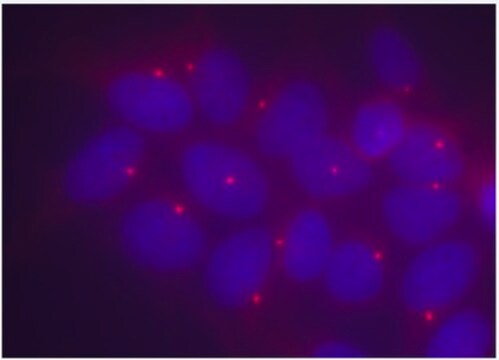
免疫細胞染色: 希釈倍率1:500で使用、HeLa細胞とA431細胞中のニネインが検出できます。
免疫沈降: 抗体は、L929細胞中のニネインを免疫沈降させることが示されています。(Delgehyr, 2005)。
免疫沈降: 抗体は、L929細胞中のニネインを免疫沈降させることが示されています。(Delgehyr, 2005)。
抗ニネイン抗体、クローン79-160-7は、WBおよびIPで使用するためのニネインに対する抗体です。
品質
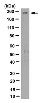
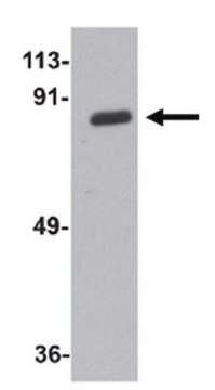
Hek293細胞ライセートのウェスタンブロットで評価されています。
ウェスタンブロッティング:1 µg/mLで使用、10 µgのHek293細胞ライセート中のニネインを検出できます。
ウェスタンブロッティング:1 µg/mLで使用、10 µgのHek293細胞ライセート中のニネインを検出できます。
ターゲットの説明
約250 kDa、実測MW。 約50 kDaに未同定の非特異的バンドが出現します。
物理的形状
0.1 M Tris-グリシン、pH 7.4、150 mM NaCl、0.05%アジ化ナトリウムを含むバッファー中の精製マウスモノクローナル IgG2aκ。
フォーマット:精製
その他情報
濃度:ロットに固有の濃度につきましては試験成績書をご参照ください。
適切な製品が見つかりませんか。
製品選択ツール.をお試しください
保管分類コード
12 - Non Combustible Liquids
WGK
WGK 1
引火点(°F)
Not applicable
引火点(℃)
Not applicable
適用法令
試験研究用途を考慮した関連法令を主に挙げております。化学物質以外については、一部の情報のみ提供しています。 製品を安全かつ合法的に使用することは、使用者の義務です。最新情報により修正される場合があります。WEBの反映には時間を要することがあるため、適宜SDSをご参照ください。
Jan Code
MABT29:
試験成績書(COA)
製品のロット番号・バッチ番号を入力して、試験成績書(COA) を検索できます。ロット番号・バッチ番号は、製品ラベルに「Lot」または「Batch」に続いて記載されています。
David K Moss et al.
Journal of cell science, 120(Pt 17), 3064-3074 (2007-08-19)
Cell-to-cell contact and polarisation of epithelial cells involve a major reorganisation of the microtubules and centrosomal components. The radial microtubule organisation is lost and an apico-basal array develops that is no longer anchored at the centrosome. This involves not only
Nathalie Delgehyr et al.
Journal of cell science, 118(Pt 8), 1565-1575 (2005-03-24)
The centrosome organizes microtubules by controlling nucleation and anchoring processes. In mammalian cells, subdistal appendages of the mother centriole are major microtubule-anchoring structures of the centrosome. It is not known how newly nucleated microtubules are anchored to these appendages. We
Ninein leads the way in vessel sprouting.
Bautch, Victoria L
Arteriosclerosis, Thrombosis, and Vascular Biology, 28, 2094-2095 (2008)
Susanne Graser et al.
The Journal of cell biology, 179(2), 321-330 (2007-10-24)
Primary cilia (PC) function as microtubule-based sensory antennae projecting from the surface of many eukaryotic cells. They play important roles in mechano- and chemosensory perception and their dysfunction is implicated in developmental disorders and severe diseases. The basal body that
Aamir Ali et al.
Nature communications, 14(1), 289-289 (2023-01-27)
Organization of microtubule arrays requires spatio-temporal regulation of the microtubule nucleator γ-tubulin ring complex (γTuRC) at microtubule organizing centers (MTOCs). MTOC-localized adapter proteins are thought to recruit and activate γTuRC, but the molecular underpinnings remain obscure. Here we show that
ライフサイエンス、有機合成、材料科学、クロマトグラフィー、分析など、あらゆる分野の研究に経験のあるメンバーがおります。.
製品に関するお問い合わせはこちら(テクニカルサービス)