
おすすめの製品


由来生物
mouse
品質水準
抗体製品の状態
purified immunoglobulin
抗体製品タイプ
primary antibodies
クローン
6D1.5/A5, monoclonal
交差性
human
テクニック
ELISA: suitable
multiplexing: suitable
western blot: suitable
アイソタイプ
IgG1
NCBIアクセッション番号
UniProtアクセッション番号
輸送温度
wet ice
ターゲットの翻訳後修飾
unmodified
遺伝子情報
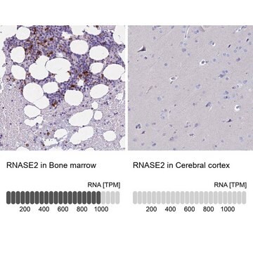
human ... RNASE2(6036)
詳細
Non-secretory ribonuclease (EC 3.1.27.5; UniProt P10153; also known as Eosinophil-derived neurotoxin, Ribonuclease 2, Ribonuclease US, RNase 2, RNase UpI-2) is encoded by the RNASE2 (also known as EDN, RAF3, RNS2) gene (Gene ID 6036) in human. The major basic protein (MBP), eosinophil cationic protein (ECP), eosinophil derived neurotoxin (EDN) and eosinophil peroxidase (EPO), are eosinophil effector molecules implicated in host defense, immunoregulatory responses, and in allergic and inflammatory reactions. These highly cationic proteins are stored preformed within the eosinophil secondary granules and released in intact membrane-bound granules during cell lysis or through a complex system of vesiculotubular structures during “piecemeal necrosis”. While eosinophil count and eosinophil surface expression of the activation marker CD69 are significantly correlated with serum concentrations of MBP, EDN and EPO, studies show that they are not correlated with ECP level. On the other hand, eosinophilic disease patients with normal eosinophil count show significantly increased concentrations of MBP, ECP, EDN, and EPO compared to normal donors. The measurement of MBP, ECP, EDN, and EPO levels thus provides a better predictor than eosinophil count alone in disease diagnosis. EDN is produced with a signal peptide sequence (a.a. 1-27), the removal of which yields the mature (a.a. 28-161) protein.
特異性
Clone 6D1.5/A5 specifically reacted with EDN, but not other granule proteins by Western blotting analysis (Makiya, M.A., et al. (2014). J. Immunol. Methods. 411:11-22).
免疫原
Purified human EDN.
アプリケーション
Research Category
炎症及び免疫
炎症及び免疫
Research Sub Category
炎症及び自己免疫疾患
炎症及び自己免疫疾患
Anti-Eosinophil-Derived Neurotoxin Antibody, clone 6D1.5/A5 is an antibody against Eosinophil-Derived Neurotoxin for use in Western Blotting, ELISA, Multiplexing.
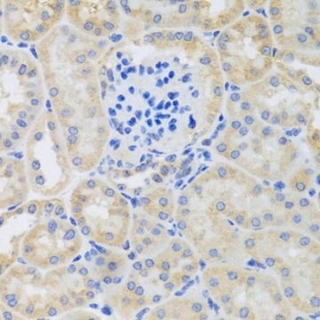
Western Blotting Analysis: 1.0 µg/mL from a representative lot detected eosinophil-derived neurotoxin in 50 µg of human liver and lung tissue lysates.
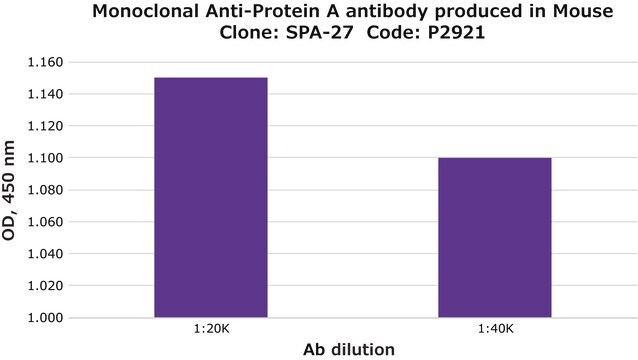
ELISA Analysis: A representative lot was employed as the capture antibody for the detection of purified human EDN, as well as EDN in serum samples from individuals with eosinophilic disorders by sandwich ELISA (Makiya, M.A., et al. (2014). J. Immunol. Methods. 411:11-22),
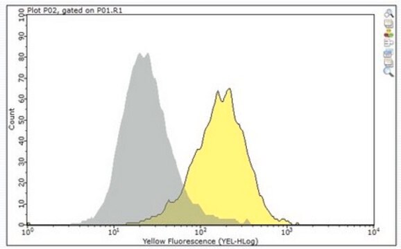
Multiplexing Analysis: A representative lot was employed as the capture antibody in a bead-based multiplex assay for the detection of purified human EDN as well as EDN in serum samples from healthy donors and individuals with eosinophilic disorders. Prior reduction and alkylation of the serum samples (by DTT and iodoacetamide, respectively) resulted in higher EDN levels (Makiya, M.A., et al. (2014). J. Immunol. Methods. 411:11-22),
ELISA Analysis: A representative lot was employed as the capture antibody for the detection of purified human EDN, as well as EDN in serum samples from individuals with eosinophilic disorders by sandwich ELISA (Makiya, M.A., et al. (2014). J. Immunol. Methods. 411:11-22),
Multiplexing Analysis: A representative lot was employed as the capture antibody in a bead-based multiplex assay for the detection of purified human EDN as well as EDN in serum samples from healthy donors and individuals with eosinophilic disorders. Prior reduction and alkylation of the serum samples (by DTT and iodoacetamide, respectively) resulted in higher EDN levels (Makiya, M.A., et al. (2014). J. Immunol. Methods. 411:11-22),
品質
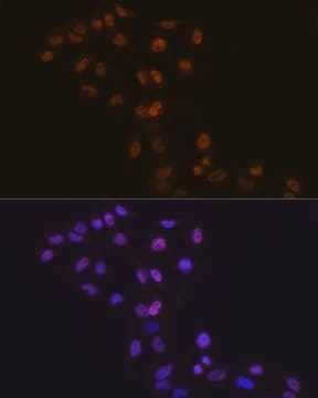
Evaluated by Western Blotting in human spleen tissue lysate.
Western Blotting Analysis: 1.0 µg/mL of this antibody detected eosinophil-derived neurotoxin in 50 µg of human spleen tissue lysate.
Western Blotting Analysis: 1.0 µg/mL of this antibody detected eosinophil-derived neurotoxin in 50 µg of human spleen tissue lysate.
ターゲットの説明
~20 kDa observed. Target band appears larger than the calculated molecular weights of 18.35 kDa (pro-form) and 15.46 kDa (mature) due to glycosylation. Uncharacterized band(s) may appear in some lysates.
物理的形状
Protein G Purified
Format: Purified
Purified mouse monoclonal IgG1 antibody in buffer containing 0.1 M Tris-Glycine (pH 7.4), 150 mM NaCl with 0.05% sodium azide.
保管および安定性
Stable for 1 year at 2-8°C from date of receipt.
その他情報
Concentration: Please refer to lot specific datasheet.
免責事項
Unless otherwise stated in our catalog or other company documentation accompanying the product(s), our products are intended for research use only and are not to be used for any other purpose, which includes but is not limited to, unauthorized commercial uses, in vitro diagnostic uses, ex vivo or in vivo therapeutic uses or any type of consumption or application to humans or animals.
適切な製品が見つかりませんか。
製品選択ツール.をお試しください
WGK
nwg
引火点(°F)
does not flash
引火点(℃)
does not flash
適用法令
試験研究用途を考慮した関連法令を主に挙げております。化学物質以外については、一部の情報のみ提供しています。 製品を安全かつ合法的に使用することは、使用者の義務です。最新情報により修正される場合があります。WEBの反映には時間を要することがあるため、適宜SDSをご参照ください。
Jan Code
MABF985:
試験成績書(COA)
製品のロット番号・バッチ番号を入力して、試験成績書(COA) を検索できます。ロット番号・バッチ番号は、製品ラベルに「Lot」または「Batch」に続いて記載されています。
ライフサイエンス、有機合成、材料科学、クロマトグラフィー、分析など、あらゆる分野の研究に経験のあるメンバーがおります。.
製品に関するお問い合わせはこちら(テクニカルサービス)