特異性
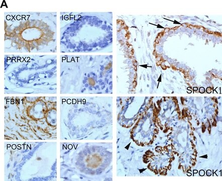
Clone 10D1 is a mouse monoclonal antibody that detects CXCR7. It targets an epitope within the extracellular domain.
免疫原
Mouse L1.2 cells transfected with human CXCR7 and stimulated for 20 h with butyric acid (5 mM).
アプリケーション
Quality Control Testing
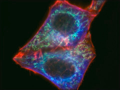
Evaluated by Flow Cytometry in Raji cells.
Flow Cytometry Analysis: 1 µg of this antibody detected CXCR7 in one million Raji cells.
Tested Applications
Inhibition Assay: A representative lot inhibited the binding to the principal ligand CXCL12 to CXCR7. (Patent ID: WO2010141986A1).
Flow Cytometry Analysis: A representative lot detected CXCR7 in Flow Cytometry applications (Patent ID: WO2010141986A1).
Note: Actual optimal working dilutions must be determined by end user as specimens, and experimental conditions may vary with the end user
ターゲットの説明
Atypical chemokine receptor 3 (UniProt: P25106; also known as C-X-C chemokine receptor type 7, CXC-R7, CXCR-7, Chemokine orphan receptor 1, G-protein coupled receptor 159, G-protein coupled receptor RDC1 homolog, RDC-1) is encoded by the ACKR3 (also known as CMKOR1, CXCR7, GPR159, RDC1) gene (Gene ID: 57007) in human. CXCR7 is a multi-pass membrane protein with four extracellular domains, seven transmembrane domains, and four cytoplasmic domains. It predominantly localized to endocytic vesicles and upon stimulation by the ligand internalizes via clathrin-coated pits in a β-arrestin-dependent manner. Once internalized, the ligand dissociates from the receptor and is targeted for degradation while the receptor is recycled back to the cell membrane. CXCR7 is an atypical chemokine receptor that controls chemokine levels and localization via high-affinity chemokine binding that is uncoupled from classic ligand-driven signal transduction cascades, resulting instead in chemokine sequestration, degradation, or transcytosis. It is also reported to act as a chemokine-scavenging receptor or chemokine decoy receptor. It acts a as a receptor for chemokines CXCL11 and CXCL12/SDF1. Chemokine binding to CXCR7 does not activate G-protein-mediated signal transduction but instead induces β-arrestin recruitment, leading to ligand internalization and activation of MAPK signaling pathway. In glioma cells, it is reported to transduce signals via MEK/ERK pathway, mediating resistance to apoptosis and promote cell growth and survival. In normal hematopoietic progenitors it is not involved in cell migration, adhesion, or proliferation. However, in malignant cells, its activation by CXCL11 leads to ERK1/2 phosphorylation that causes enhanced cell adhesion and migration. (Ref.: Wang, C., et al. (2018). Front. Pharmacol. 9; 641; Rajagopal, S., et al. (2010). Proc.Natl. Acad.Sci. USA. 107(2); 628-632; Levoye, A., et al. (2009). Blood. 113(24); 6085-6093; Zabel, BA., et al. (2009). J. Immunol. 183(5); 3204-3211).
物理的形状
Purified mouse monoclonal antibody IgG2a in PBS without azide.
再構成
0.5 mg/mL. Please refer to guidance on suggested starting dilutions and/or titers per application and sample type.
保管および安定性
Store at -10°C to -25°C. Handling Recommendations: Upon receipt and prior to removing the cap, centrifuge the vial and gently mix the solution. Aliquot into microcentrifuge tubes and store at -20°C. Avoid repeated freeze/thaw cycles, which may damage IgG and affect product performance.
その他情報
Concentration: Please refer to the Certificate of Analysis for the lot-specific concentration.
免責事項
Unless otherwise stated in our catalog or other company documentation accompanying the product(s), our products are intended for research use only and are not to be used for any other purpose, which includes but is not limited to, unauthorized commercial uses, in vitro diagnostic uses, ex vivo or in vivo therapeutic uses or any type of consumption or application to humans or animals.