
MABE286
Anti-Replication Protein A Antibody, clone RPA34-19
clone RPA34-19, from mouse
別名:
Replication protein A 32 kDa subunit, RP-A p32, Replication factor A protein 2, RF-A protein 2, Replication protein A 34 kDa subunit, RP-A p34
ログイン組織・契約価格を表示する
すべての画像(3)
About This Item
UNSPSCコード:
12352203
eCl@ss:
32160702
NACRES:
NA.41
おすすめの製品
由来生物
mouse
品質水準
抗体製品の状態
purified antibody
抗体製品タイプ
primary antibodies
クローン
RPA34-19, monoclonal
交差性
human
テクニック
immunocytochemistry: suitable
immunohistochemistry: suitable
western blot: suitable
アイソタイプ
IgG1κ
NCBIアクセッション番号
UniProtアクセッション番号
輸送温度
wet ice
ターゲットの翻訳後修飾
unmodified
遺伝子情報
human ... RPA2(6118)
詳細
RPA (RP-A p32) is a heterotrimeric protein complex that binds specifically to single-stranded DNA (ssDNA). It is composed of three subunits: RPA1 (70 kDa), RPA2 (32 kDa), and RPA3 (14 kDa). RPA plays multiple roles in DNA replication. At the onset of DNA replication, RPA is loaded onto chromatin, and is needed for subsequent loading of DNA polymerase and other replication proteins to initiate DNA replication. After replication begins, RPA moves with replication forks, stabilizing ssDNA and assisting in DNA synthesis. In addition to its replication function, RPA is also known to play essential roles in damage repair and recombination. The 32 kDa subunit is phosphorylated by the cdc2 family of kinases when cells enter S-phase; and by ATM, ATR, and DNA-PK proteins in response to DNA damage.
免疫原
Replication Protein A purified from U293 cells.
アプリケーション
Research Category
エピジェネティクス及び核内機能分子
エピジェネティクス及び核内機能分子
Research Sub Category
細胞周期、DNA複製及び修復
細胞周期、DNA複製及び修復
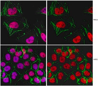
Anti-Replication Protein A Antibody, clone RPA34-19 is a Mouse Monoclonal Antibody for detection of Replication Protein A also known as Replication protein A 32 kDa subunit, RP-A p32 & has been validated in WB, ICC & IHC.
Immunocytochemistry Analysis: A 1:250 dilution from a reprsentative lot detected Replication Protein A in A431 cells.
Immunohistochemistry Analysis: A 1:5 dilution from a representative lot detected Replication Protein A in human placental chorionic villi and in human colorectal adenocarcinoma tissue.
Immunohistochemistry Analysis: A 1:5 dilution from a representative lot detected Replication Protein A in human placental chorionic villi and in human colorectal adenocarcinoma tissue.
品質
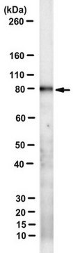
Evaluated by Western Blot in HeLa cell lysate.
Western Blot Analysis: A 1:2,000 dilution of this antibody detected Replication Protein A in 10 µg of HeLa cell lysate.
Western Blot Analysis: A 1:2,000 dilution of this antibody detected Replication Protein A in 10 µg of HeLa cell lysate.
ターゲットの説明
~34 kDa observed. This protein has 3 isoforms: Isoform 1 (~29 kDa), Isoform 2 (~30 kDa), and Isoform 3 (~39 kDa).
物理的形状
Protein G Purified
Format: Purified
Purified mouse monoclonal IgG1κ in buffer containing 0.1 M Tris-Glycine (pH 7.4), 150 mM NaCl with 0.05% sodium azide.
保管および安定性
Stable for 1 year at 2-8°C from date of receipt.
アナリシスノート
Control
HeLa cell lysate
HeLa cell lysate
免責事項
Unless otherwise stated in our catalog or other company documentation accompanying the product(s), our products are intended for research use only and are not to be used for any other purpose, which includes but is not limited to, unauthorized commercial uses, in vitro diagnostic uses, ex vivo or in vivo therapeutic uses or any type of consumption or application to humans or animals.
適切な製品が見つかりませんか。
製品選択ツール.をお試しください
保管分類コード
12 - Non Combustible Liquids
WGK
WGK 1
引火点(°F)
Not applicable
引火点(℃)
Not applicable
適用法令
試験研究用途を考慮した関連法令を主に挙げております。化学物質以外については、一部の情報のみ提供しています。 製品を安全かつ合法的に使用することは、使用者の義務です。最新情報により修正される場合があります。WEBの反映には時間を要することがあるため、適宜SDSをご参照ください。
Jan Code
MABE286:
試験成績書(COA)
製品のロット番号・バッチ番号を入力して、試験成績書(COA) を検索できます。ロット番号・バッチ番号は、製品ラベルに「Lot」または「Batch」に続いて記載されています。
Motohiro Yamauchi et al.
DNA repair, 7(3), 405-417 (2008-02-06)
Several DNA damage checkpoint factors form nuclear foci in response to ionizing radiation (IR). Although the number of the initial foci decreases concomitantly with DNA double-strand break repair, some fraction of foci persists. To date, the physiological role of the
Janna Luessing et al.
iScience, 25(7), 104536-104536 (2022-06-28)
Abscission, the final stage of cytokinesis, occurs when the cytoplasmic canal connecting two emerging daughter cells is severed either side of a large proteinaceous structure, the midbody. Here, we expand the functions of ATR to include a cell-cycle-specific role in
ライフサイエンス、有機合成、材料科学、クロマトグラフィー、分析など、あらゆる分野の研究に経験のあるメンバーがおります。.
製品に関するお問い合わせはこちら(テクニカルサービス)